Figure 1. SLAT/Def6 deficient mice
are inferior to their WT controls in developing EAU. A:
Disease scores of individual mice of the two mouse lines.
Horizontal lines are mean ±SEM. A summary of repeated
experiments comparing the response in mice of the two lines. **
p<0.005. B: Sections of representative eyes of the
two groups. The changes in the eye of the control mouse are
markedly more severe and mainly include infiltration of
inflammatory cells in various ocular tissues, as well as retinal
folding and loss of photoreceptor cells (score: 2+). Only minor
infiltration is seen in the SLAT/Def6 deficient mouse eye
(score: 0.5).
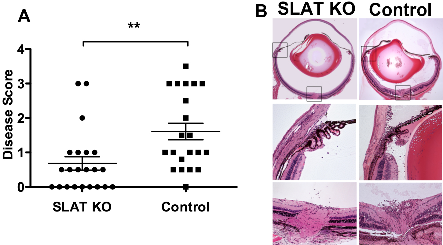
 Figure 1
of Vistica, Mol Vis 2012; 18:1858-1864.
Figure 1
of Vistica, Mol Vis 2012; 18:1858-1864.