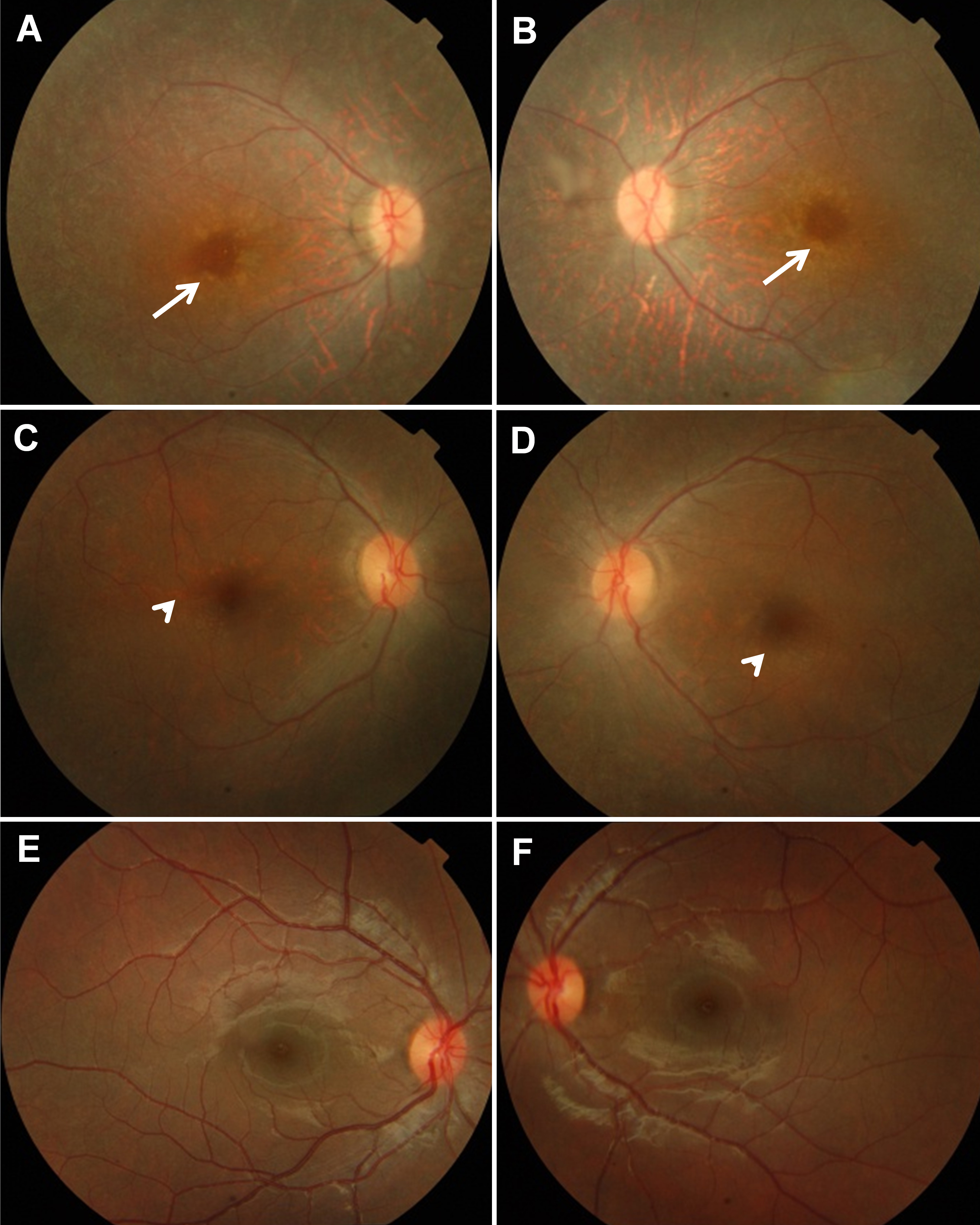
Figure 2. Fundus photographs of
affected individuals from both families and of a normal
individual.
A,
B: Right and left fundus,
respectively, of the proband of family A (see arrow,
Figure 1A),
representative of the fundus appearance of all affected members
of this family. Arrows indicate yellow perifoveal annular rings.
C,
D: Right and left fundus, respectively, of the
proband of family B (see arrow,
Figure 1B). Arrowheads
point to the developing perifoveal annular rings.
E,
F:
Right and left fundus, respectively, of a normal individual.
 Figure 2
of Ajmal, Mol Vis 2012; 18:1226-1237.
Figure 2
of Ajmal, Mol Vis 2012; 18:1226-1237.