Figure 1. Pedigrees and TULP1
genotyping results for families A and B. A: This is the
pedigree of family A in which the presence of the c.1138A>G
variant (M) was found in a homozygous state in all affected
individuals. As expected for causal autosomal recessive
variants, unaffected parents are heterozygous, and normal
individuals carry one or no mutant allele. B: This is
the pedigree of family B in which the presence of the
c.1445G>A variant (M) was shown in a homozygous state in the
6 affected individuals, and heterozygously in an unaffected
sibling, parents and grandparents of affected persons. C:
This is the sequence electropherogram showing the nucleotide
change from adenine to guanine in the proband of family A (upper
panel), and sequence electropherogram of a control individual
showing the wild-type adenine (lower panel). D: This is
the sequence electropherogram of the proband carrying the mutant
adenine in family B (upper panel); and the sequence
electropherogram of a control individual with the normal guanine
(lower panel). Probands are indicated with arrows; asterisks
indicate the individuals that were tested using HumanOmniExpress
(>700K) SNP microarrays. M/M, homozygous c.1138A>G (in
family A) or homozygous c.1445G>A (in family B); +/M,
heterozygous mutations present; +/+, two wild-type alleles
present.
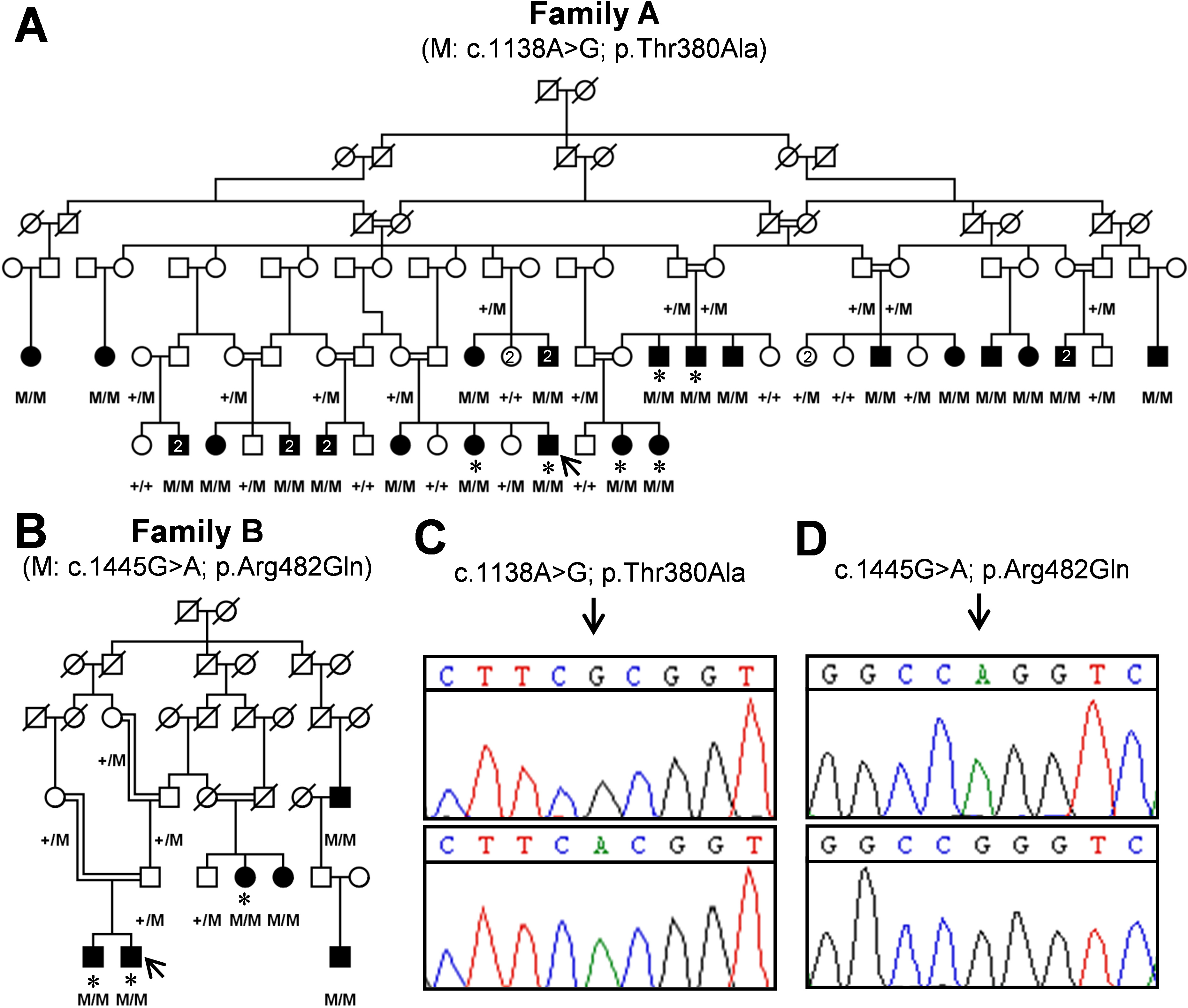
 Figure 1
of Ajmal, Mol Vis 2012; 18:1226-1237.
Figure 1
of Ajmal, Mol Vis 2012; 18:1226-1237.