Figure 1. Human corneal endothelial
cell examination. Inverted phase-contrast imaging (A) and
immunofluorescent ZO-1 staining (B) of the human corneal
endothelial cells. Human corneal endothelial cells were observed
in mosaic pattern. Immunofluorescence with anti-ZO-1 antibody, a
general marker of the tight junction, revealed the morphology.
The nuclei were stained using Hoechst 33342 (blue).
Magnification, 200×.
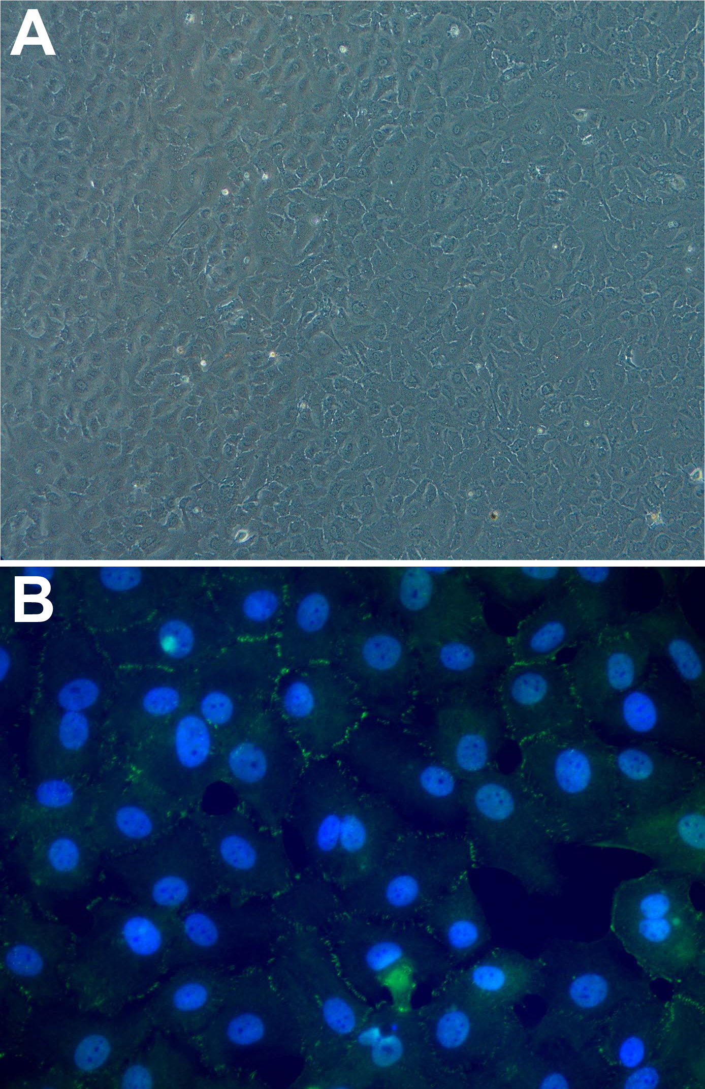
 Figure 1
of Shin, Mol Vis 2011; 17:3371-3378.
Figure 1
of Shin, Mol Vis 2011; 17:3371-3378.