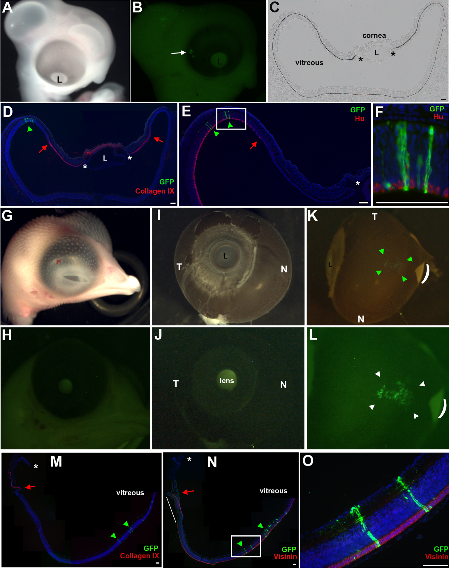
Figure 3. Non-optic cup lip
derivatives populate the posterior neural retina.
A-
E:
The optic cup was targeted with retrovirus, as described in
Figure 1E,
and reincubated until E5 (
A).
B: Whole mount view
of GFP expressing cells (arrow), located at a distance from the
OCL.
C,
D: Transverse sections through the GFP
expressing region. GFP+ cells do not populate the ciliary body
(Collagen IX signal) or the margin between the ciliary body and
neural retina (red arrow). Asterisks indicate OCL.
E,
F:
GFP+ cells are found only in the Hu+ neural retina. Boxed region
in
E is represented at higher magnification in
F.
G-
O: Results of optic cup labeling, examined at
E10.
G,
H: GFP+ cells are not immediately
detectable in whole mount view of head.
I,
J:
Infected eye dissected from head, nasal (N) and temporal (T)
orientation as indicated. No GFP signal is seen in anterior view
of eye.
K,
L: GFP+ cells are seen at the
posterior-dorsal aspect of the optic cup (bracketed by green
arrowheads). Curved line indicates a tear in the RPE.
L:
Slightly higher magnification of
K.
M-
O:
Sections through eye in
K. GFP+ cells are only found in
the posterior retinal domain (green arrowheads). The ora
serrate/CMZ, defined as the junction between the ciliary body
(Collagen IX signal,
M) and neural retina (Visinin
signal,
N), is indicated by the red arrow. GFP+ cells
are not found at the CMZ. Asterisk indicates OCL. Note, isolated
eye tissue is delicate and does not adhere well upon sectioning.
In panel N the tissue has flipped during processing (white
line). Boxed are in N shown at higher magnification in
O.
Scale Bars: 100 µm. N; Nasal, T; temporal, L; Lens.
 Figure 3
of Venters, Mol Vis 2011; 17:3347-3363.
Figure 3
of Venters, Mol Vis 2011; 17:3347-3363.