Figure 1. Optic cup labeling reveals
a precise growth pattern for cells derived from the optic cup
lip. A, B: Graphics of the labeling targets in
the E3 optic cup, viewed as a wholemount (A) and
transverse section (B). The crosshairs in A
highlight orientation in all wholemount eyes: D-dorsal;
V-ventral; N-nasal; T-temporal. In B, A-anterior (from
lens), P-posterior (toward head) axis, specific for the eye, is
indicated. The OCL (green dot) or optic cup (purple dot) was
targeted around the circumference of the pupil. C-M: DiI
labeled embryos: Embryos were imaged immediately after labeling
(C-E) and following reincubation (F-M).
The curved bars outline the boundary between the lens and optic
cup. C, D: Dye targeted to the OCL (arrowhead).
E: Dye targeted on the optic cup (arrowhead). F,
G: OCL targeted embryos after 24 (F) and 48 (G)
h reincubation. Labeled cells are present as a spoke-line
extending from the OCL, adjacent to the lens, toward the
posterior eye. The line highlights the extent of labeled cells.
H, I: Resultant dye pattern 24 (H) and 48
(I) h after labeling the optic cup, as in E.
Discrete patches of dye are found posterior to the OCL (arrow).
The scattered dye is in the mesenchyme overlying the
neuroepithelium. No linear labeling is found. J-M:
Sections through the labeled tissue 48 h after targeting the OCL
(J, K) or optic cup (L, M). J:
After OCL labeling, the dye is retained in the OCL and is
distributed through the anterior optic cup. K: A higher
magnification of the boxed area in J. Dye is present in
the inner and outer epithelial layers of the optic cup and in
the overlying mesenchyme. L: After optic cup labeling,
dye is found as a discrete patch in the posterior eye and is
absent from the front of the eye. Some dye is present in the
mesenchyme overlying the neuroepithelium (arrowheads). M:
A higher magnification of the boxed area in L. Dye is
present in both the neural retina and overlying retinal
pigmented epithelium. Scale Bars: 100 µm. OCL; Optic Cup Lip, L;
lens, NR; Neural retina, RPE; Retinal pigmented epithelium, CB;
Ciliary body, IL; Inner layer, OL; Outer layer.
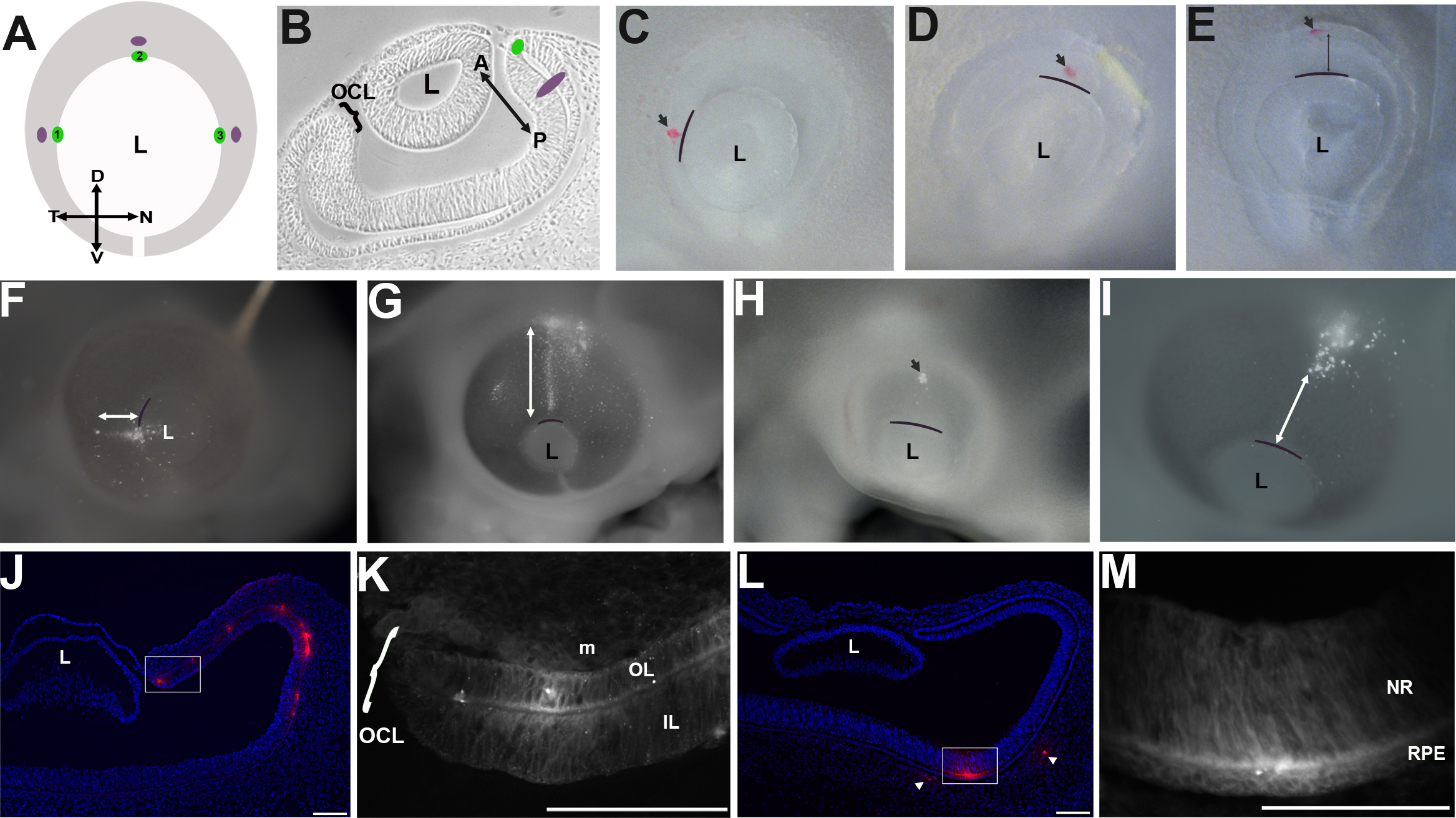
 Figure 1
of Venters, Mol Vis 2011; 17:3347-3363.
Figure 1
of Venters, Mol Vis 2011; 17:3347-3363.