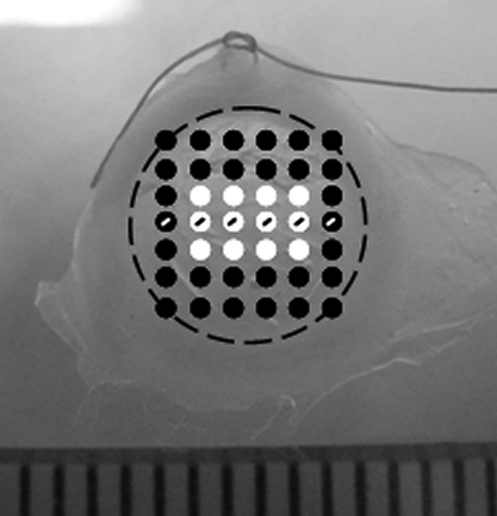
Figure 1. Small-angle X-ray scatter
patterns (represented by circles) were collected at 1 mm
intervals over each guinea pig cornea. Twelve measurements of
fibril diameter, fibril separation distance, and D-periodicity
from the central cornea were used (white circles) to produce
weighted averages for each treatment group (
Table 1).
Measurements across the horizontal meridian of each cornea
(circles containing a line) were averaged within treatment
groups to show limbus-to-limbus changes in fibril separation
distance and fibril diameter (
Figure 3). The position of
the limbus is shown as a dashed black line. A scleral suture at
the 12 o’clock position ensured that in vivo corneal orientation
was maintained during data collection.
 Figure 1
of Hayes, Mol Vis 2011; 17:3107-3115.
Figure 1
of Hayes, Mol Vis 2011; 17:3107-3115.