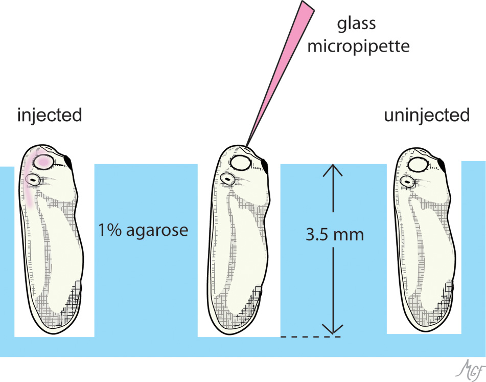
Figure 2. This diagram illustrates the position of embryos in agarose dishes for brain ventricle injections. Embryos were placed in
foxhole arrays punched out of 1% agarose dishes (blue). At least 25 foxholes can be prepared per dish. The holes are made
to a depth so that the head protrudes slightly out of the foxhole (see Methods). The embryonic diencephalic ventricle is located
along the midline, and protrudes anteriorly just under the frontal ectoderm and prosencephalon (compare with
Figure 3). A midline anterior approach conveniently accesses the ventricle without disturbing the optic vesicles (middle embryo).
The injected material (pink) readily fills the brain ventricle with optic vesicle, and typically can be appreciated extending
into the central canal [
30].
 Figure 2 of
Gonzalez-Fernandez, Mol Vis 2011; 17:2956-2969.
Figure 2 of
Gonzalez-Fernandez, Mol Vis 2011; 17:2956-2969.