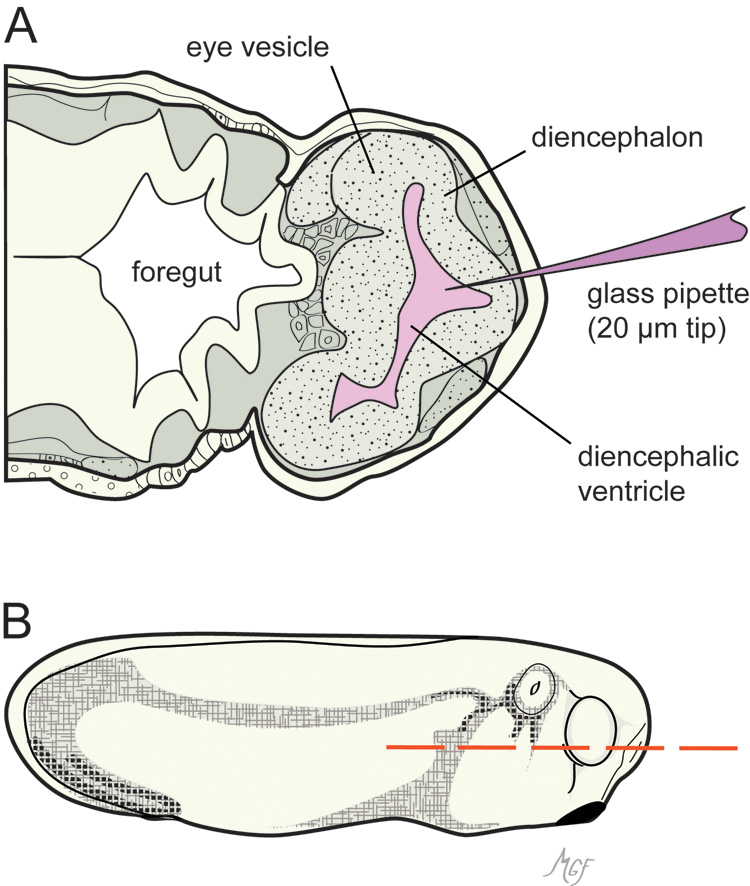
Figure 3. Horizontal section of a stage 26 embryo illustrating the brain ventricle injection method used to introduce materials into
the optic vesicle lumen.
A: A glass micropipette was introduced into the diencephalic ventricle anteriorly.
B: The dashed line shows the orientation of the section in panel
A. The drawings were prepared based on [
24,
30].
 Figure 3 of
Gonzalez-Fernandez, Mol Vis 2011; 17:2956-2969.
Figure 3 of
Gonzalez-Fernandez, Mol Vis 2011; 17:2956-2969.