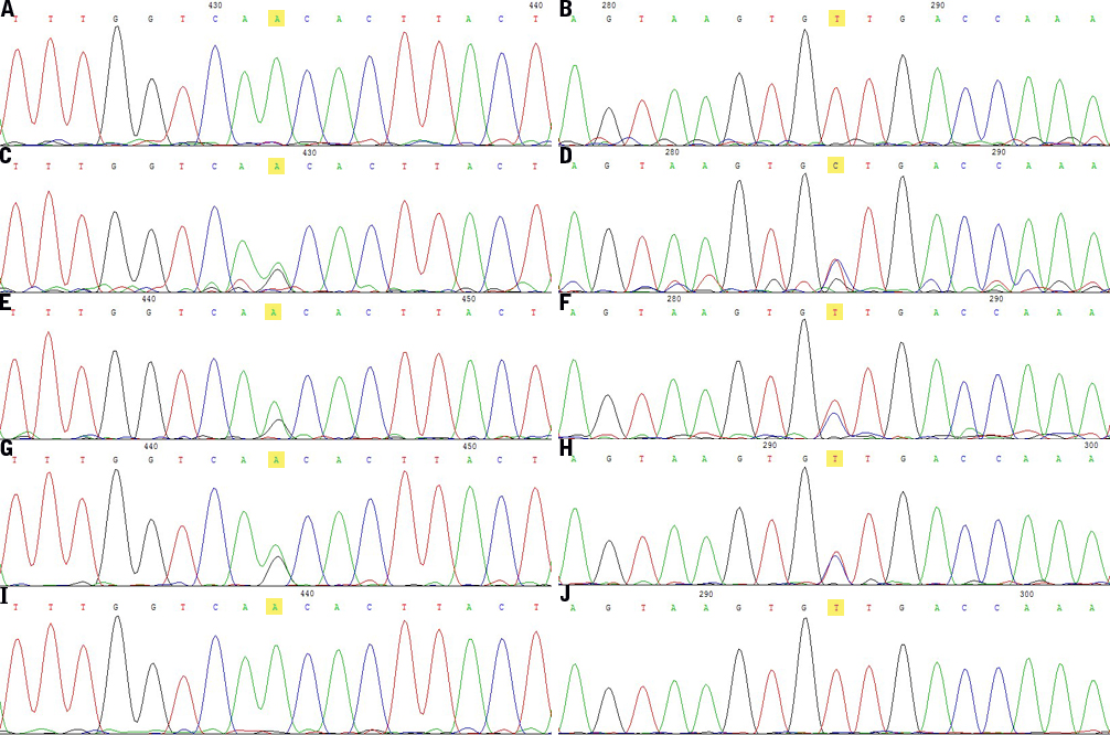
Figure 5. Mutation in
UBIAD1. Chromatograms of the subjects whose DNA samples were sequenced directly showing heterozygous mutation N102S in exon 1. Normal
sequence of
UBIAD1 near codon 102 detected in a healthy control (
A,
B) is showed on the first of forward and reverse reading, respectively. The left ones are forward reading, and the right ones
are reverse reading. The sequence in the proband (
C,
D) shows a heterozygous A>G transversion (at condon 102 leads to a change from asparagine (AAC) to serine (AGC), which is highlighted
in yellow), which is also in other affected members: the proband’s daughter (
E,
F), and the proband’s granddaughter (
G,
H), but not in any unaffected members or normal controls. The indeterminate phenotype member (
I,
J) in
Figure 1 is determined as unaffected individual due to the chromatograms are the same with the controls (
A,
B).
 Figure 5 of
Du, Mol Vis 2011; 17:2685-2692.
Figure 5 of
Du, Mol Vis 2011; 17:2685-2692.