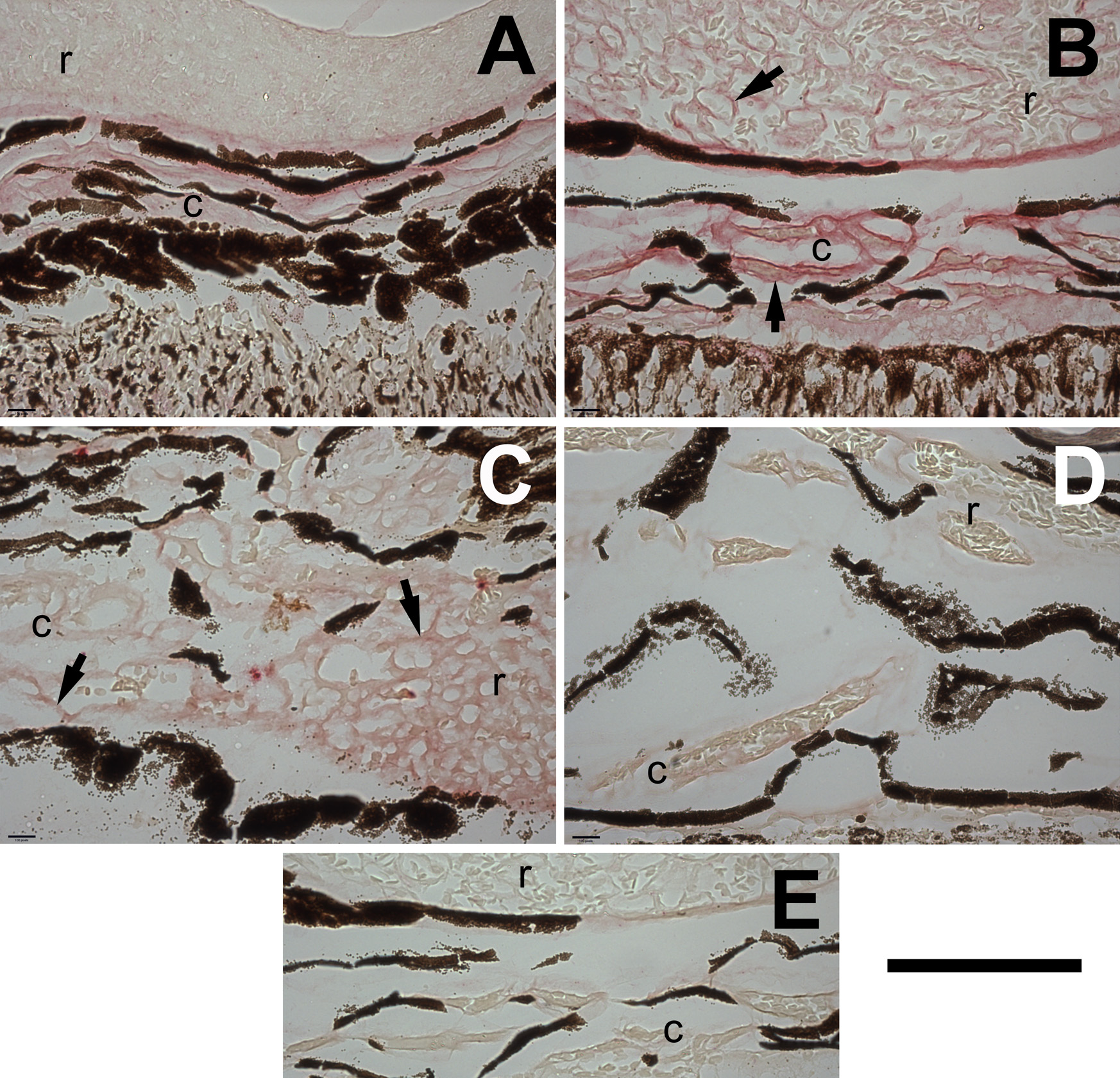
Figure 4. Tbdn and ZO-1 proteins show
reciprocal regulation in cold-adapted smelt rete and choroidal
blood vessels. Compared to warm fish maintained at 8–10 °C
(
A: warm specimen/Tbdn stain;
C: warm
specimen/ZO-1 stain), the endothelial linings of the choroidal
(c) and rete (r) blood vessels of cold fish maintained at
0.5 °C show a higher expression level of Tbdn protein, but
a lower level of ZO-1 protein in these regions (
B: cold
specimen/Tbdn stain;
D: cold specimen/ZO-1 stain). The
arrows indicate choroidal and rete blood vessel endothelia.
E:
Sections were also incubated with a control IgG and showed no
staining of the blood vessels. Positive staining for Tbdn (OE5)
and ZO-1 appears as bright red staining. The dark brown or black
color in all panels is intrinsic due to pigmentation from the
pigments cells of the choroidal vasculature and/or retinal
tissue. The immunohistochemical results shown here are
representative of three smelt in each of the warm and cold
groups and are quantitated in
Figure 6. The scale bar
in the lower right corner of the figure indicates 100 μm.
 Figure 4
of Gendron, Mol Vis 2011; 17:2596-2604.
Figure 4
of Gendron, Mol Vis 2011; 17:2596-2604.