Figure 6. Quantitation of staining by
a mouse anti-Tbdn antibody (A) and by a mouse anti-ZO-1
antibody (B) on smelt eye tissues. Compared to fish
maintained at 8–10 °C (warm), there was a significant
increase (p<0.001; n=15 color intensity measurements per
group) in Tbdn staining of rete-choroidal blood vessels in the
cold-adapted smelt (maintained at 0.5 °C [cold]), as shown
in panel A. There was no significant difference in the
staining of Tbdn in the neural retina. Compared to fish
maintained at 8–10 °C (warm), there was a significant
decrease (p<0.001; n=15 color intensity measurements per
group) in ZO-1 staining of rete- choroidal blood vessels in the
cold-adapted smelt (maintained at 0.5 °C [cold]), as shown
in panel B. There was no significant difference in the
staining of ZO-1 in the cornea.
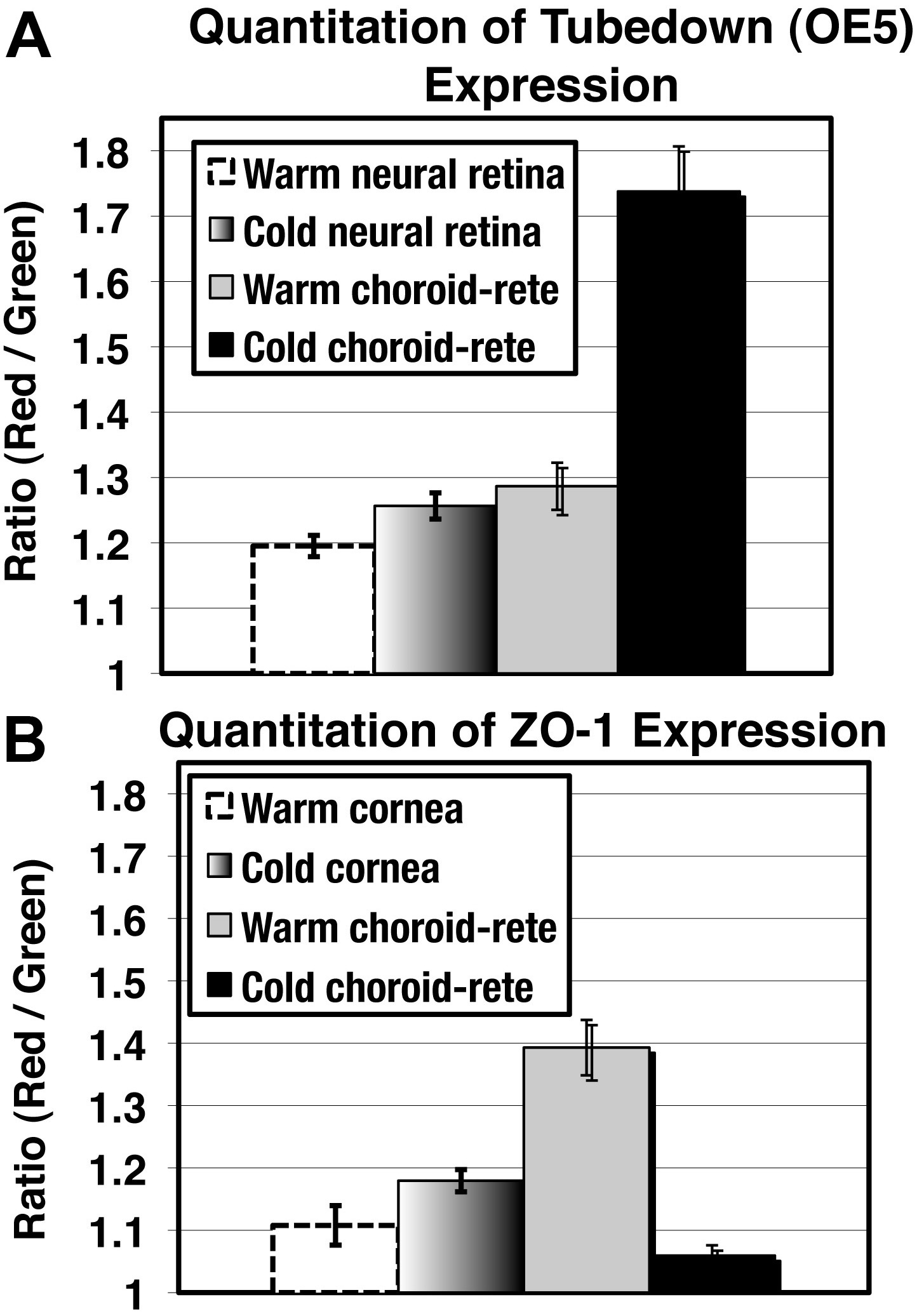
 Figure 6
of Gendron, Mol Vis 2011; 17:2596-2604.
Figure 6
of Gendron, Mol Vis 2011; 17:2596-2604.