Figure 6. Canolol activated the ERK pathway in ARPE-19 cells. ARPE-19 cells were treated with canolol (200 μM), U0126 (10 μM), or t-BH
(150 μM). A: After 1 h exposure to t-BH, a western blot analysis was performed using p-ERK and ERK antibodies. β-Actin was used as an
internal control for sample normalization. The figures were selected as representative data from three independent experiments.
A quantitative analysis was performed by measuring intensity relative to the untreated control. B: After 24 h exposure to t-BH, cell viability was measured by an MTT assay. Data was expressed as a percentage of the control.
Each value represents the mean±SEM (n=3) of three independent experiments. The single asterisk indicates p<0.05 and the double
asterisk indicates p<0.01.
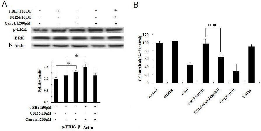
 Figure 6 of
Dong, Mol Vis 2011; 17:2040-2048.
Figure 6 of
Dong, Mol Vis 2011; 17:2040-2048.