Figure 4. Canolol inhibited t-butyl hydroxide (t-BH)-induced apoptosis in ARPE-19 cells. A: The ARPE-19 cells were incubated with 100 μM and 200 μM of canolol for 24 h, and were then treated with 150 μM of t-BH for
6 h. A flow cytometric analysis was used to quantify the rate of cell apoptosis using double staining of Annexin V-FITC and
PI. Each diagram represents three independent experiments. B: Quantitative analyses of the apoptosis rate in ARPE-19 cells. The results were represented by a mean±SEM (n=3). Data was
expressed as a percentage of the untreated control. The double asterisk indicates p<0.01 versus t-BH-induced cells without
canolol pretreatment (t-test).
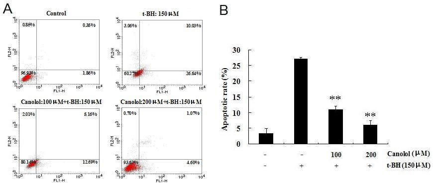
 Figure 4 of
Dong, Mol Vis 2011; 17:2040-2048.
Figure 4 of
Dong, Mol Vis 2011; 17:2040-2048.