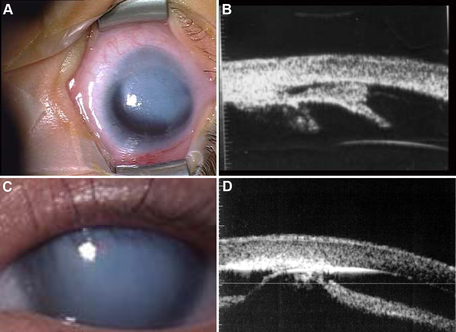
Figure 1. Iridocorneal and
keratolenticular adhesions. Peters anomaly (PA) classically describes a
congenital corneal opacity resulting from an iridocorneal (
A,
B)
or
keratolenticular adhesion (
C,
D). The opacity may be
central (
C,
D) and may take up a small part or the whole
of the cornea, or may be eccentric (
A,
B). It is
unreliable to ascribe this term judging only from the clinical picture
without making use of ultrasonography, notably ultrasound biomicroscopy
(UBM;
B,
D), which helps visualize the structures of
the anterior segment and prove or disprove the presence of adhesions (
B,
D). The use of UBM in
A and
B shows contact
between the cornea and iris (iridocorneal adhesion) and in
C
and
D contact between the cornea and the anterior lenticular
surface (keratolenticular adhesions), resulting in disorganization and
loss of clarity in the central cornea, confirming the diagnosis of PA.
NB. These opacities are avascular (see
Figure 3) and in these cases the
lens is of a normal size.
 Figure 1 of Mataftsi, Mol Vis 2011; 17:1624-1640.