Figure 3. Total corneal opacification. Two
typical examples of the phenotype designated as total congenital
corneal opacification. S-CCO (A-D). This condition
confusingly, has been previously described clinically as sclerocornea
in the literature. However, anterior segment imaging using UBM shows
that the lens has failed to form normally in case A and B
and that in case C and D there is failure of the lens
(L) to separate from the cornea (ILA) and there is an abnormal zonular
ciliary complex (Z); this suggests another primary lens problem leading
to a secondary CCO. Note both opacities are vascularized.
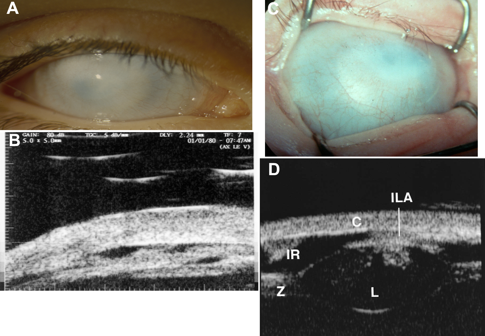
 Figure 3 of Mataftsi, Mol Vis 2011; 17:1624-1640.