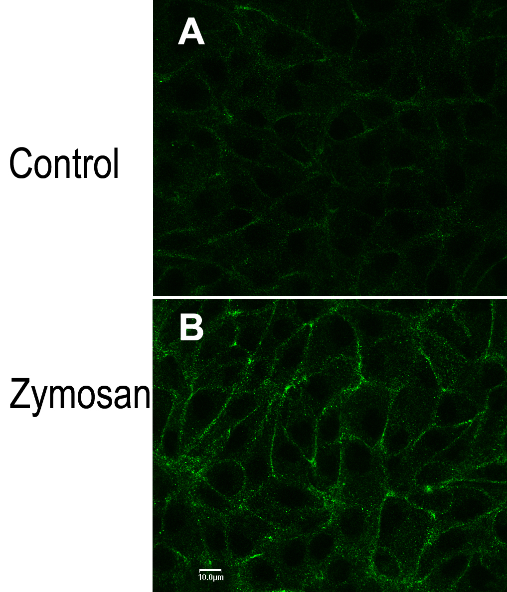
Figure 5. Transient receptor potential
vanilloid channel, member 6 (TRPV6) localization to the plasma membrane
increased after treatment with zymosan, a reagent that is phagocytized
by the retinal pigment epithelium. Donor R cells were plated (passage
151) onto Permanox four-well chamber slides and grown for 7 days under
standard culture conditions. Monolayers were rinsed free of media with
standard buffer and equilibrated in that buffer for 30 min at
37 °C. Monolayers were exposed to either standard buffer (Control,
upper panel) or standard buffer plus 2 mg/ml zymosan (Zymosan, lower
panel) for a further 10 min at 37 °C. Reaction was stopped by
formaldehyde fixation, and immunocytochemistry was performed. Primary
and secondary antibodies were those used in
Figure 4, bottom panel (TRPV6).
Nuclei were stained with TO-PRO-3. The scale bar in panel
B
represents 10 μm and applies to both panels.
 Figure 5 of Kennedy, Mol Vis 2010; 16:665-675.