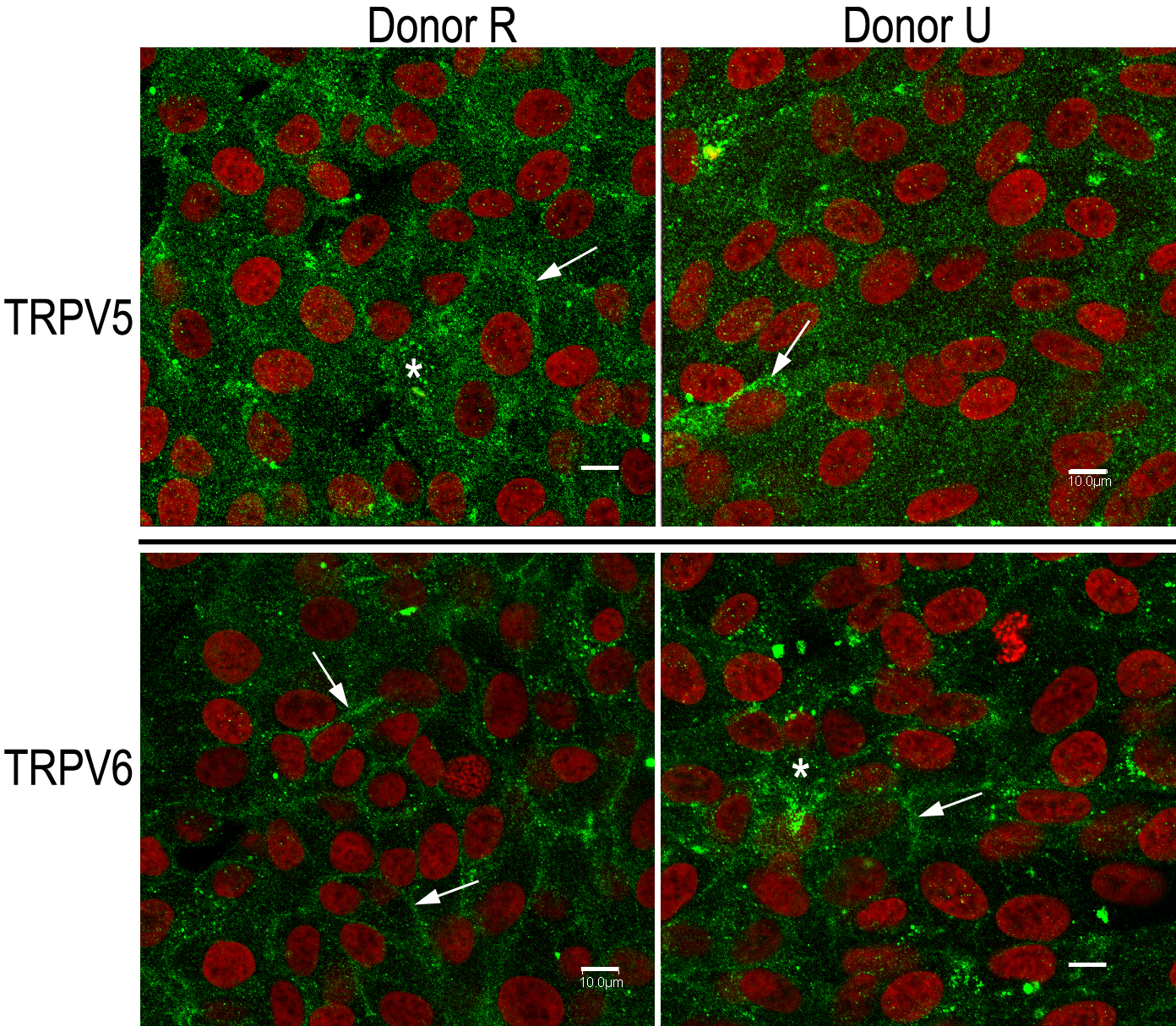
Figure 4. Immunoreactivity against
transient receptor potential vanilloid channel member 5 (TRPV5) and
member 6 (TRPV6), respectively is present on the plasma membrane as
well as in the cytoplasm of cultured adult human retinal pigment
epithelium. Donor cultures (R on the left and U on the right) are the
same as those used for western blotting (
Figure 2). Cells were plated
(passage 146 for R and passage 116 for U) onto uncoated four-well glass
chamber slides and grown for 3 days under standard culture conditions
(described in the Methods). Monolayers were stained for TRPV5 (upper
panels) using an affinity-purified, polyclonal, anti-TRPV5 antibody
(8515, final concentration 5 µg/ml). Monolayers were stained for TRPV6
(lower panels) using an affinity-purified, polyclonal, anti-TRPV6
antibody (E-16, final concentration 2 µg/ml). The same secondary
antibody was used with both primary antibodies: antigoat Alexa 488
(final dilution 1:500). Nuclei (red) were visualized with TO-PRO-3
(final concentration 2 µM). Scale bars in all panels represent 10 µm.
Both plasma membrane (arrows) and prominent, punctuate, cytoplasmic
(asterisk) staining is seen for both antibodies.
 Figure 4 of Kennedy, Mol Vis 2010; 16:665-675.