Figure 2. Western blot analysis of
transient receptor potential vanilloid channel member 5 (TRPV5) and
member 6 (TRPV6) protein expression in cultured human retinal pigment
epithelium. A: Reactivity with anti-TRPV5 polyclonal antibody
8515 (final concentration 1 µg/ml). The secondary antibody was antigoat
immunoglobulin G (IgG) conjugated to alkaline phosphatase (final
dilution 1:5,000). Samples were from three different human primary
cultures: R established from a 72-year-old Caucasian male and used at
passage 72; U established from an 82-year-old Caucasian male and used
at passage 42; W established from a 47-year-old Caucasian female and
used at passage 16. B: Reactivity with an anti-TRPV6
affinity-purified polyclonal antibody 11A (final concentration 0.5
µg/ml). The secondary antibody was antirabbit IgG conjugated to
alkaline phosphatase (final dilution 1:5,000). In B, culture R
was used at passage 83, culture U at passage 53, and culture W at
passage 27.
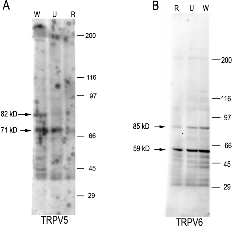
 Figure 2 of Kennedy, Mol Vis 2010; 16:665-675.