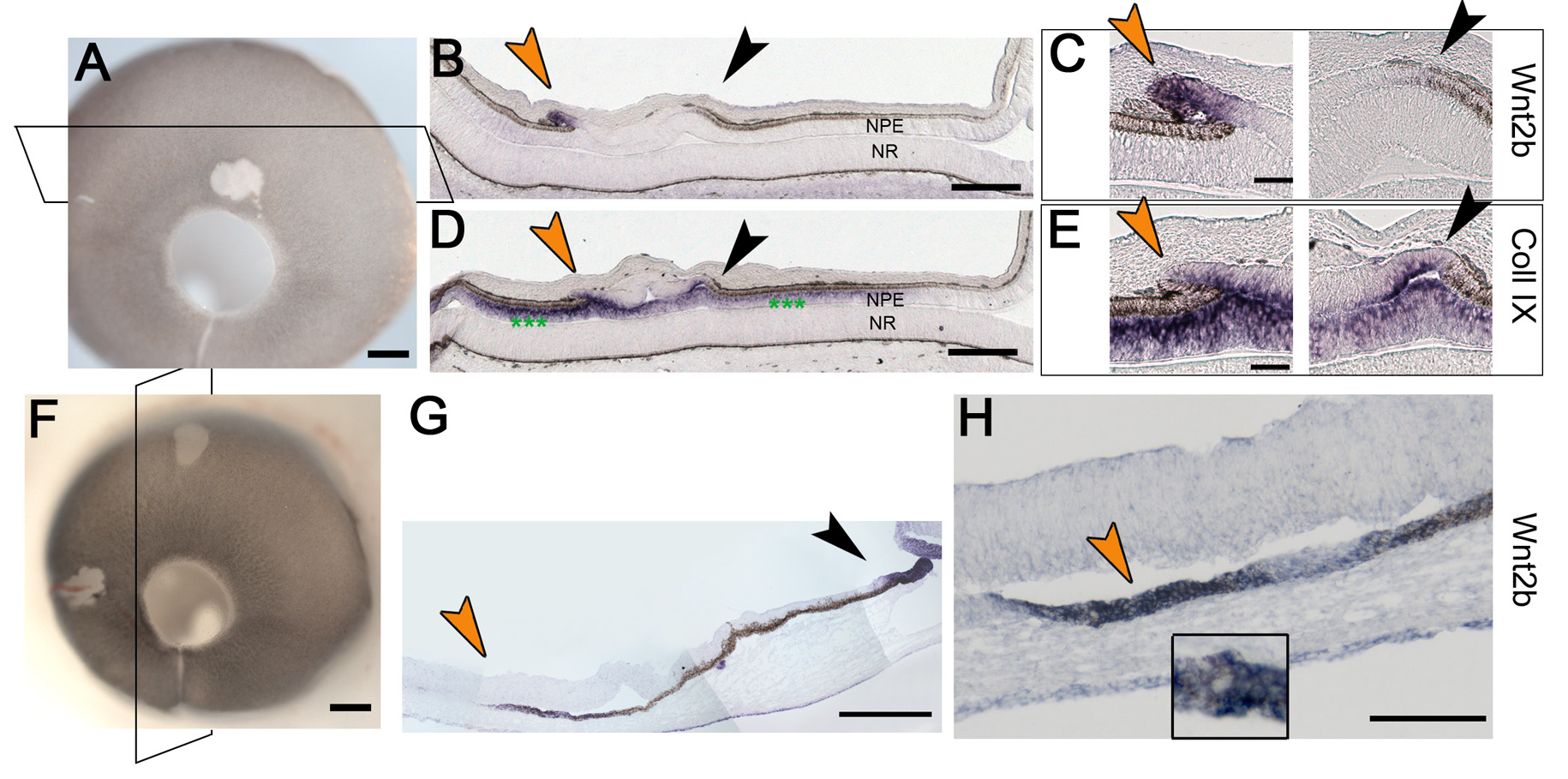
Figure 5. Wnt 2b is ectopically expressed
in transition zones
A: An infected eye at Hamburger-Hamilton
(HH) stage 25 with virus associated depigmented patch at a distance of
approximately 200 μm from lens. The section plane is indicated by a
box.
B-
E: are sections through the eye in
A.
Note that in these sections the eye is flattened and the vitreous space
is not obvious. The posterior neural retina (NR) lies directly in
contact with the anterior epithelia (NPE, nonpigmented epithelium of
the ciliary body; see
Figure 1).
B: Wnt2b gene
expression is found at the edge of the patch; arrows bracket the patch
of depigmented tissue with a transition zone at each edge.
C: A
Higher magnification view of portions of
B, showing Wnt2b in
one of the two transition zones (orange arrows) at the edges of white
patch.
D: Collagen IX expression in the section adjacent
B;
green
asterisks the indicate endogenous expression in anterior
epithelium (NPE).
E: A higher magnification view showing
collagen IX expression in both transition zones.
F: An infected
eye at HH stage 25 with a virus associated depigmented patch at a
distance of approximately 1200 microns from lens. The section plane is
indicated by a box.
G: A section through the infected area and
lens, showing endogenous Wnt2b signal in anterior optic cup lip (purple
arrow) and ectopic Wnt2b signal at edge of infected patch (orange
arrow); green asterisk indicates endogenous lens signal.
H: A
higher magnification of the transition zone in
G (orange
arrow). The inset panel shows the juxtaposition of Wnt2b expression and
pigmented epithelium. Scale bars in
A and
F are equal
to 500 µm, in
B and
D are equal to 200 µm, in
C,
E
and
H are equal to 100 µm and in
G are equal to 300
µm.
 Figure 5 of Kitamoto, Mol Vis 2010; 16:2701-2717.