Figure 1. Wnt2b is a marker of the anterior optic cup lip. A: A graphic of the chick optic vesicle at Hamburger-Hamilton (HH) stage 11 (embryonic day 1.5) in which the presumptive neural
retina (NR) is indicated in green and the presumptive retinal pigment epithelium (RPE) in brown and purple arrowheads mark
the presumptive ciliary body/iris. B: A graphic of the optic cup, formed after invagination of the optic vesicle. At HH stage 25 (embryonic day 5), the ciliary
body/iris is molecularly identifiable (light green dots, purple arrowheads). C-G: Whole mount in situ hybridization for Wnt2b gene expression through various stages of eye development. C: At HH stage 15 (embryonic day 2), a few hours after the initiation of invagination, Wnt2b expression is enriched in the
dorsal optic tissue (arrow). D: Sections through an HH stage 19 embryo (embryonic day 2.5) showing Wnt2b expression throughout the RPE (arrow) and the overlying
surface ectoderm (asterisk). The lens is outlined. E: At HH stage 21 (embryonic day 3) Wnt2b is refined to the anterior optic cup lip (arrowheads). Expression is also found throughout
the surface ectoderm (asterisk). F: At HH stage 23 (embryonic day 4) Wnt2b expression is robust in the optic cup lip, with faint expression in RPE. G: At HH stage 25 (embryonic day 5) Wnt2b is expressed exclusively in the optic cup lip (arrow); white asterisk indicates a
rip artifact, double white asterisk indicates non-specific background staining. H: Section in situ analysis confirms the restriction of Wnt2b signal (green arrow) to optic cup lip at HH stage 25. Brown is
the endogenous color of the RPE. The lens epithelium (green asterisk) and the surface ectoderm (black asterisk) also express
Wnt2b. I: Adjacent section stained for collagen IX shows the extent of specified ciliary body tissue (green arrowheads) in the anterior
of the optic cup. Dashes outline the lens. Scale bars in C-G are equal to 500 µm, and in H are equal to 100 µm. Abbreviations used are as follows: L-lens; staging table; NPE-non-pigmented epithelium of the ciliary
body.
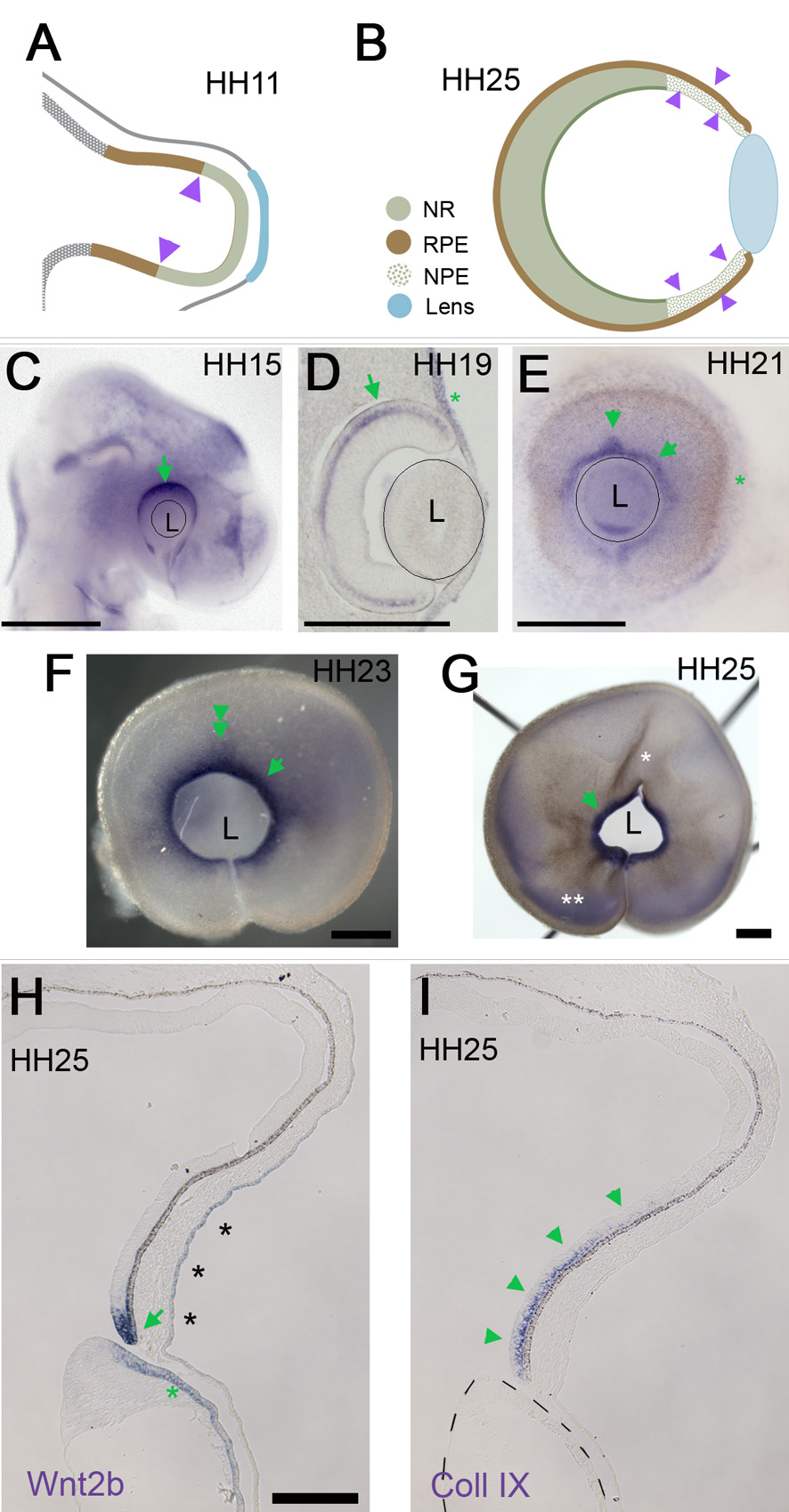
 Figure 1 of
Kitamoto, Mol Vis 2010; 16:2701-2717.
Figure 1 of
Kitamoto, Mol Vis 2010; 16:2701-2717.