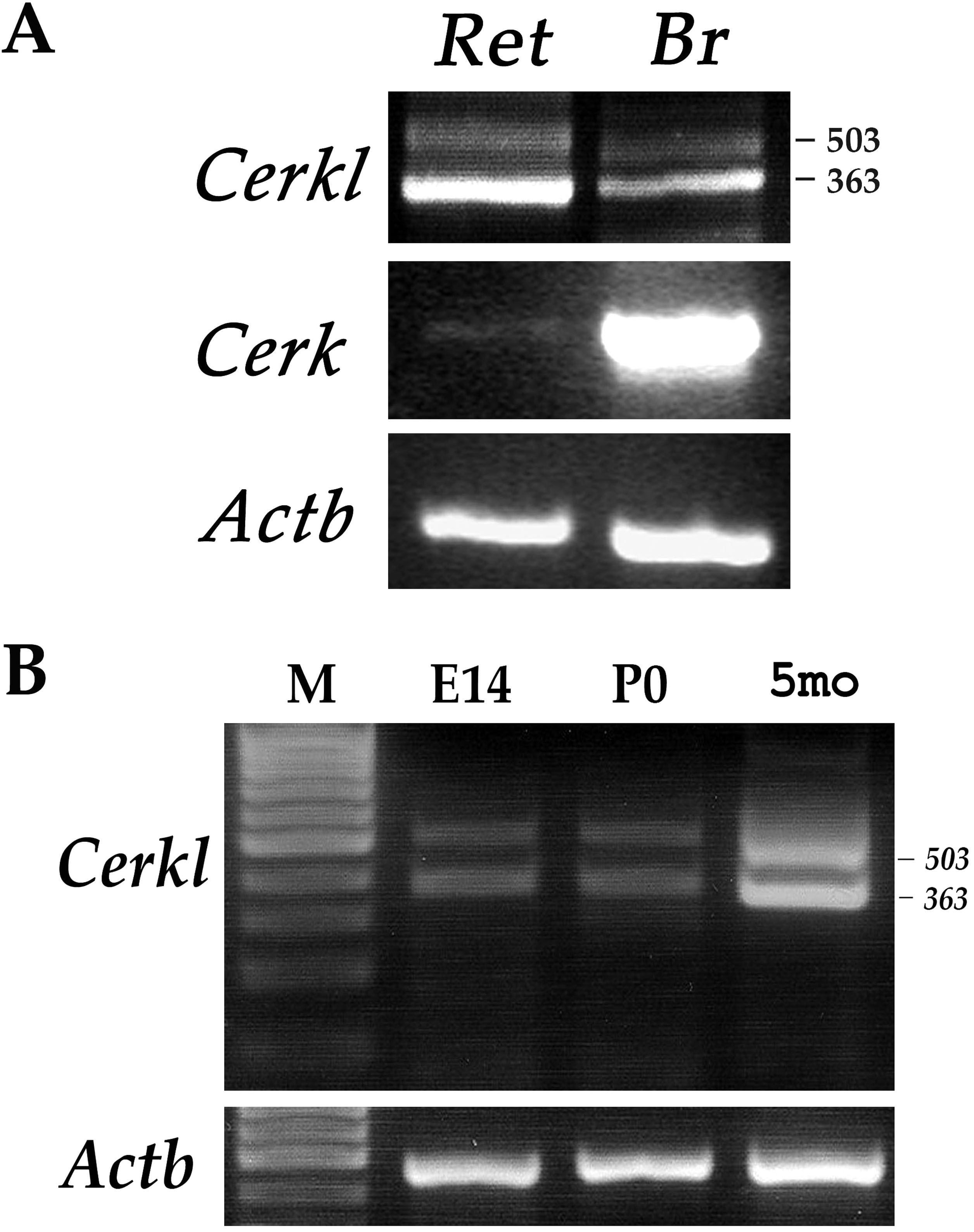
Figure 1. Expression analysis of
Cerkl
in the mouse eye.
A: Reverse Transcription-Polymerase Chain
Reaction (RT–PCR) analysis of
Cerk (412 bp) and
Cerkl
expression in mouse brain (Br) and retina (Ret).
Cerkl
expression was tested using primers located in exons 1 and 2 (see
Figure 2B).
The
two
PCR products obtained for
Cerkl (503 and 363 bp)
represent different splice-isoforms.
B: RT–PCR analysis of
Cerkl
in the mouse eye at different developmental time points: embryonic day
14 (E14), newborn (P0), and 5 months (5 mo). The analysis indicates
Cerkl expression at all time points tested. β-actin (
Actb;
437
bp
product) served as an internal control for RNA quality and
quantity. M: size marker.
 Figure 1 of Vekslin, Mol Vis 2010; 16:2539-2549.