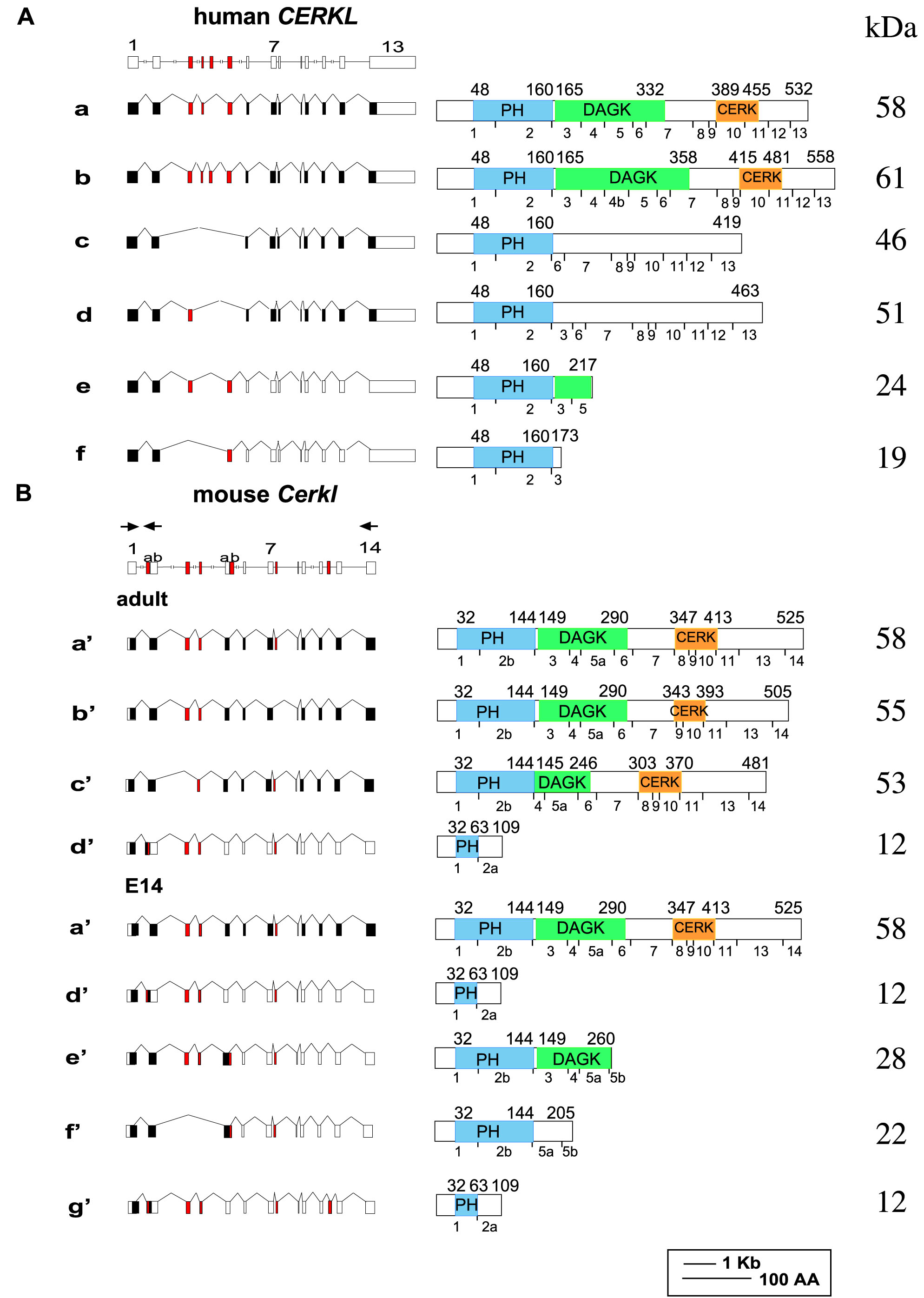
Figure 2. CERKL gene and
splice-variants in human and mouse retina. A schematic representation
of
CERKL genes (drawn to scale), splice-variants (left panels),
and expected protein products (right panels) is shown. In the
splice-variants illustrations, filled boxes represent coding exons, and
open boxes represent non-coding exons. Alternatively spliced exons are
marked in red. In the protein products illustrations, the PH, DAGK, and
CERK homology domains are indicated. The borders of each domain are
marked by their amino acid positions, indicated above each protein. The
breaks between exons are indicated below each protein. Protein
molecular weight in kDa is shown on the right.
A: Human
CERKL
gene and splice-variants previously identified in the human retina.
Variants e and f correspond to
AY690333 and
AY690332 [
13].
B: Mouse
Cerkl
gene and splice-variants found in the mature and embryonic (E14) mouse
retina. Two of the mouse isoforms (isoforms d’ and g’) encode for the
same short protein product. Mouse isoform a’, which is present in both
adult and embryonic samples, is equivalent to human isoform a, and
mouse isoform f’ is equivalent to human isoform f. Locations of PCR
primers used for RT–PCR analysis and for splice-variant identification
(forward primer located in exon 1 and reverse primers located in exons
2 and 14) are indicated by arrows above the schematic representation of
the murine gene.
 Figure 2 of Vekslin, Mol Vis 2010; 16:2539-2549.