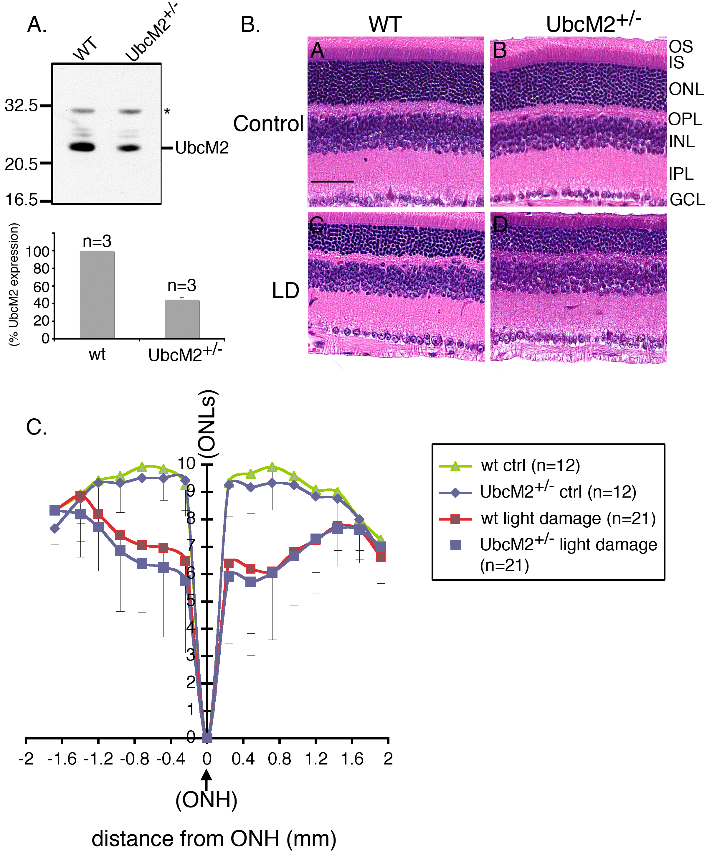
Figure 4. Mice harboring a single intact
allele of UbcM2 are not more susceptible to light-induced retinal
degeneration.
A: Inactivation of a single UbcM2 allele reduces
expression of the enzyme by 58%. Equal amounts (10 μg) of retinal
lysates derived from a UbcM2
+/− mouse and a wild-type
(WT) littermate were subjected to denaturing and reducing sodium
dodecyl sulfate PAGE (SDS–PAGE) followed by anti-UbcM2 western
blotting. The migration of molecular weight markers is shown on the
left. The asterisk denotes a nonspecific band serving as a loading
control. The graph depicts the relative level of expression of UbcM2 in
WT versus heterozygous littermates (n=3 of each genotype) as determined
using a desktop scanner and Image J software.
B: Representative
hematoxylin and eosin (H&E) stained paraffin-embedded sections are
shown from mice (UbcM2
+/− and WT littermates) 7 days
after acute bright-light challenge. Control denotes animals that were
maintained in dim light for the entire experiment. Abbreviations of
retinal cell layers are as described in the legend for
Figure 3.
The magnification bar in panel A represents 50 μm.
C: A Spider
graph representing data compiled from the indicated number of animals
for each experimental condition. The error bars represent the standard
deviation. Outer nuclear layer (ONL) rows are plotted along the
y-axis
with
inferior and superior distances in mm from the optic nerve head
(ONH) depicted along the
x-axis. There was no statistically
significant difference in ONLs between WT and UbcM2
+/−
control animals or between WT and UbcM2
+/−
light-damaged animals.
 Figure 4 of Mirza, Mol Vis 2010; 16:2425-2437.