Figure 3. Differential expression of class
III ubiquitin conjugating enzymes (E2s) in cells of the mouse retina. A-I:
Shown
here are representative photomicrographs of paraffin-embedded
sections labeled with the indicated antibodies. Mouse age in weeks is
shown in the bottom right corner of each panel. Abbreviations are as
follows: RPE represents retinal pigment epithelium; OS represents outer
segments; IS represents inner segments; ONL represents outer nuclear
layer; OPL represents outer plexiform layer; INL represents inner
nuclear layer; IPL represents inner plexiform layer; GCL represents
ganglion cell layer. The magnification bar represents 50 μm, and all
photomicrographs were taken at the same magnification. J-R:
Enlarged sections are shown of images from (J) highlighting the
presence or absence of RPE labeling with the various antibodies. Black
arrowheads mark unlabeled RPE nuclei and white arrows indicate nuclei
labeled with anti-UbcM2. The magnification bar represents 25 μm.
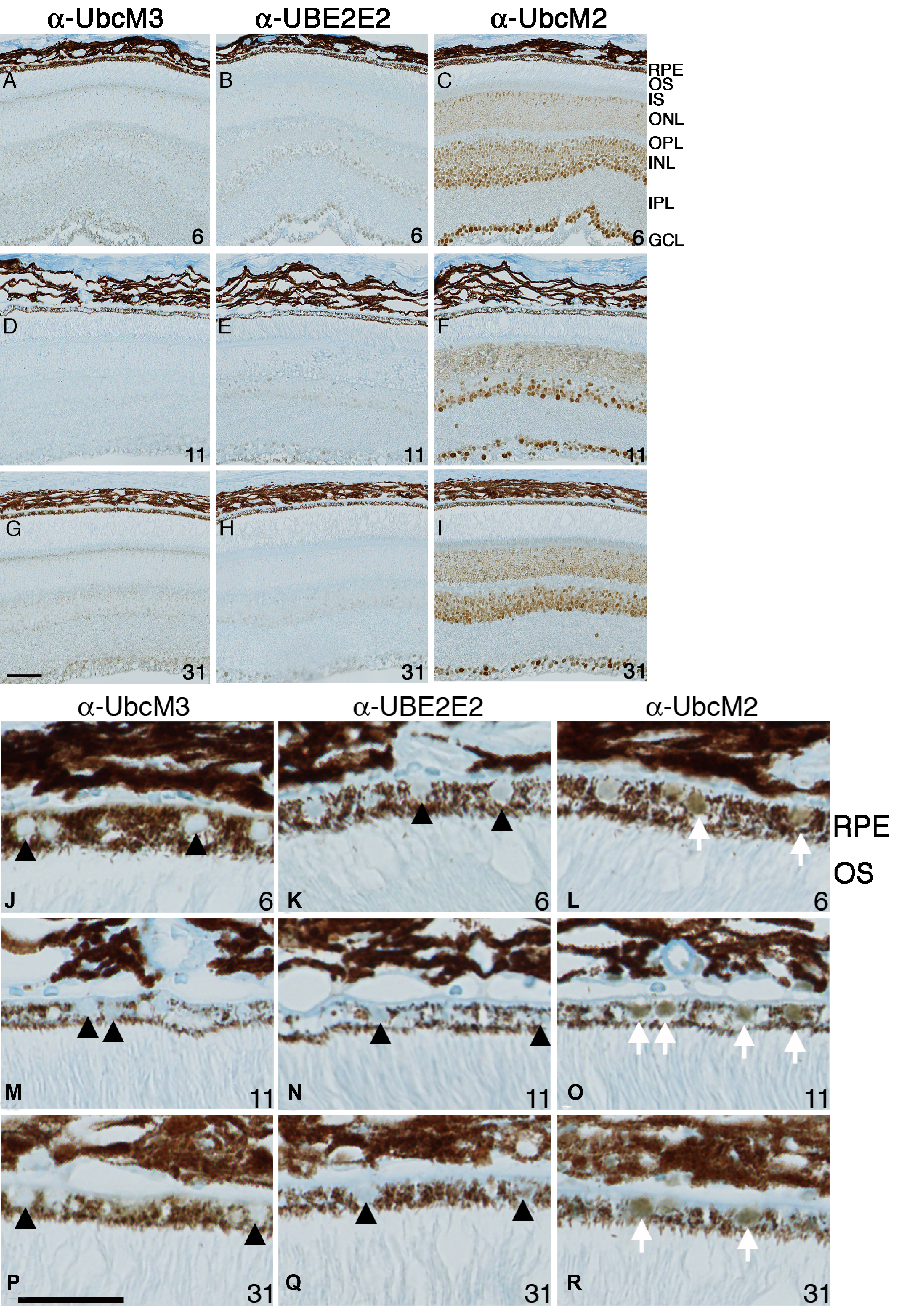
 Figure 3 of Mirza, Mol Vis 2010; 16:2425-2437.