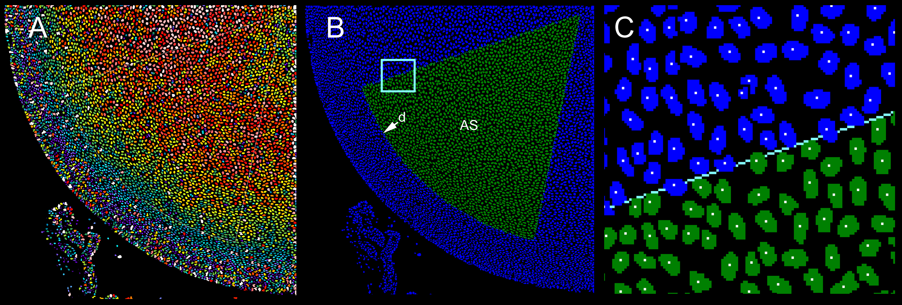
Figure 5. Computer-assisted identification
and quantification of nuclei in a 60° sector (AS) of the anterior lens
epithelium.
A: Nuclei are identified using the count nuclei
application. Colors indicate nuclear size (large nuclei are shown in
warmer colors).
B: Following image segmentation, nuclei lying
within sector AS are counted (green). The arc of the sector is located
at d, the fiduciary nucleus (see
Figure 3 and
Figure 4).
C: A higher magnification view of the boxed area in
B.
The position of the centroid (center of mass) of each nucleus is
calculated (indicated by a white dot). Only nuclei with centroids
located within AS are included in the analysis.
 Figure 5 of Bassnett, Mol Vis 2010; 16:2294-2300.