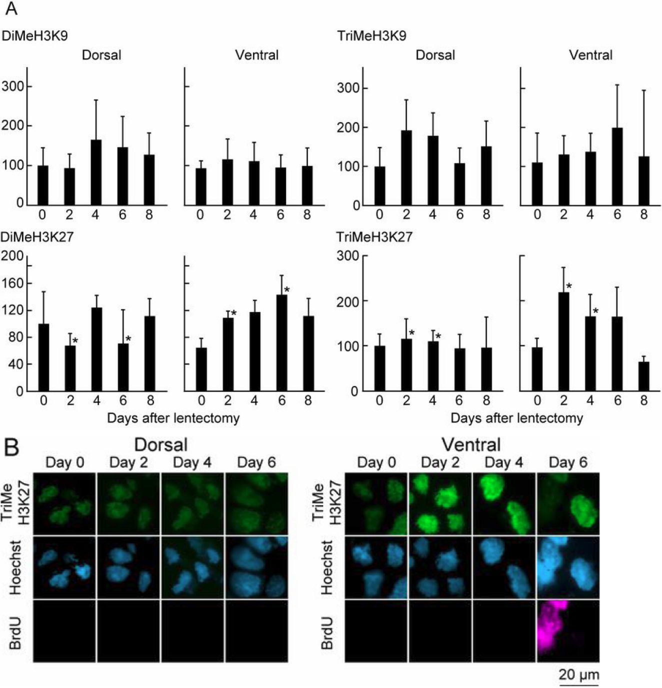
Figure 2. Changes in histone modifications
related to gene repression during early lens regeneration. Same
procedure as in
Figure
1.
A: Quantification of detected signal by
immunohistochemistry using histone modification antibodies.
B:
Immunohistochemistry using TriMeH3K27 antibody, showing patterns in
dorsal and ventral iris at different time points.
 Figure 2 of Maki, Mol Vis 2010; 16:1893-1897.