Figure 1. Changes in histone modifications related to gene activation during early lens regeneration. A: Quantification of detected signal by immunohistochemistry using histone modification antibodies. To examine the cells before
cell cycle reentry, BrdU was administrated everyday. Immunohistochemistry was performed and the detected signal intensity
of each histone modification in nucleus of PECs without BrdU incorporation was measured. The signal intensity of histone modification
was normalized with that of Hoechst 33528. The value of normalized signal in dorsal iris on day 0 is represented as 100%.
Error bars, standard deviation (n=5–17). Asterisks indicate a significant difference at p<0.03, Student’s t test (2-tailed) between dorsal and ventral iris at same day. B: Immunohistochemistry. Staining patterns in dorsal iris are shown at different time points.
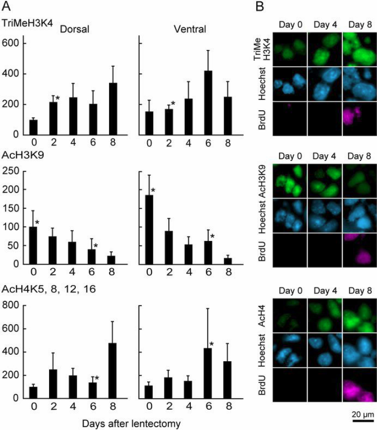
 Figure 1 of
Maki, Mol Vis 2010; 16:1893-1897.
Figure 1 of
Maki, Mol Vis 2010; 16:1893-1897.