Figure 1 of
Yan, Mol Vis 2010; 16:1736-1742.
Figure 1.
Histological examination of left biceps from the patient.
A
: H&E staining shows an RRF (arrow).
B
: MGT staining shows an atypical RRF (arrow).
C
: SDH staining shows ragged-blue fibers (arrows). Magnification 400×.
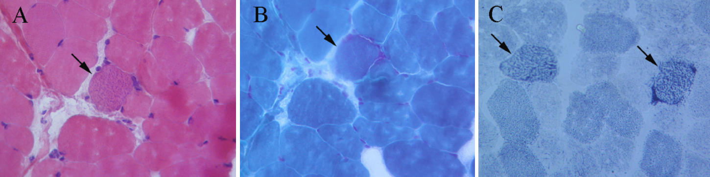
 Figure 1 of Yan, Mol Vis 2010; 16:1736-1742.