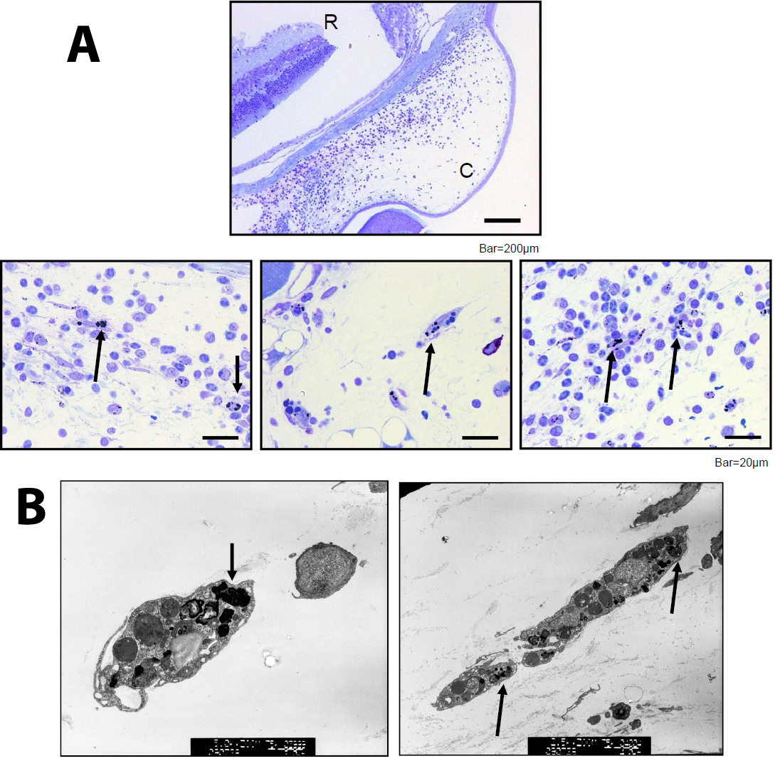
Figure 3. Examination of cells that engulfed OVA-gold in the conjunctiva. Conjunctival samples were prepared as described in
Figure 2. Cells that engulfed OVA-gold were examined histologically by light (
A) and electron microscopy (
B). Gold particles were detected in the cytoplasma of cells in the conjunctiva (
A). C and R indicates conjunctiva and retina, respectively (
A). Gold particles were present in cytoplasms of oval-shaped cells and spindle-like-shaped cells (
B). Arrows indicate cells that engulfed gold particles.
 Figure 3 of
Ishida, Mol Vis 2010; 16:1280-1285.
Figure 3 of
Ishida, Mol Vis 2010; 16:1280-1285.