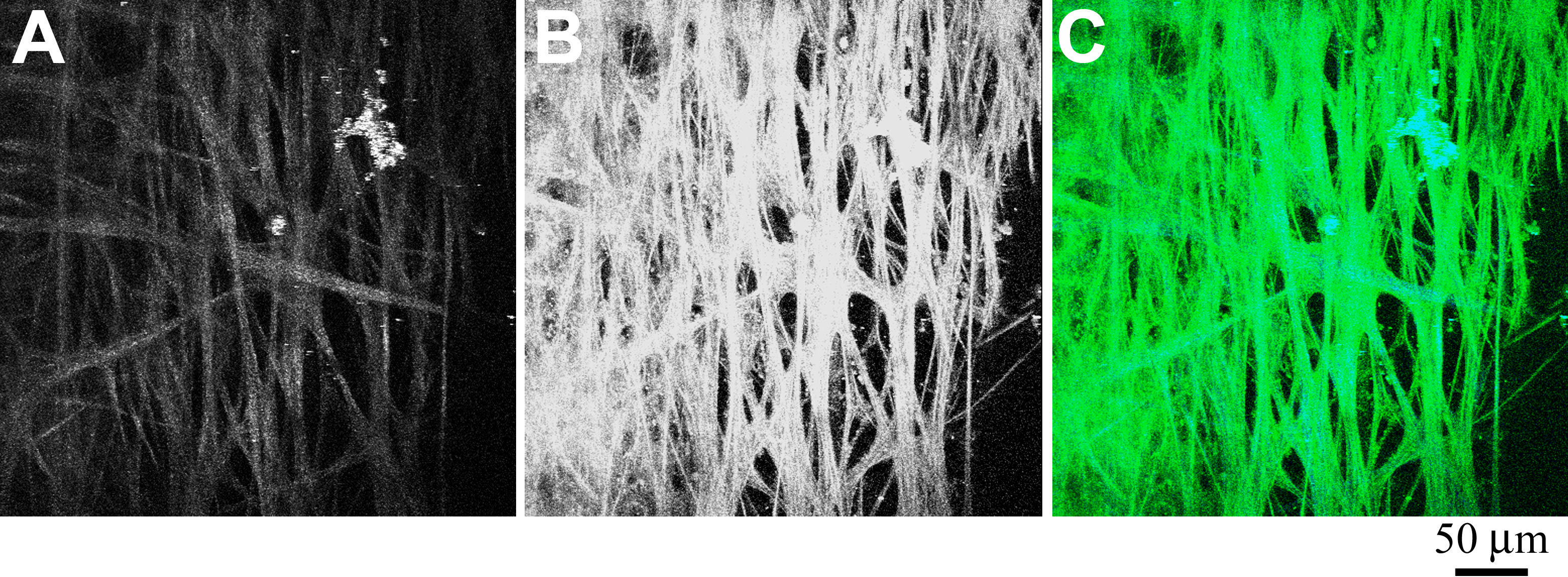
Figure 5. Second-harmonic generation (SHG)
and AF of TM region of a human eye from a 73-year-old donor. A section
of a human eye was flat-mounted with the anterior chamber facing the
microscope objective. Images represent a projection of the multiple
z-sections flattened into a single plane.
A: The SHG emission
(388 nm to 409 nm) collected from an 800 nm excitation of TM.
B:
AF
collected simultaneously as described in
Figure 1.
C: Merged image of
SHG (blue) and AF (green) emission. Black scale bar=50 µm.
 Figure 5 of Ammar, Mol Vis 2010; 16:935-944.