Figure 1. Autofluorescence (AF) of the
cornea/TM region of a human eye from a 73-year-old donor. A section of
a tissue was flat-mounted with the anterior chamber facing the
microscope objective. An AF window (452 nm to 644 nm) was collected
from an 800 nm excitation. 100 z-sections were imaged at 1 micron
intervals using a 25× objective. A: A flattened projection of
all z-sections at of the junction of cornea/TM. The curvature of the
tissue where the cornea and TM meet (top of A) places it beyond
the working distance of the objective lens, and therefore it appears as
a dark region. B: A higher resolution image of the TM region. C:
Image
snapshots of a 3D reconstruction of the cornea/TM region, rotated
around the y-axis, shown at intervals of 30°. White/black scale
bars=100 µm.
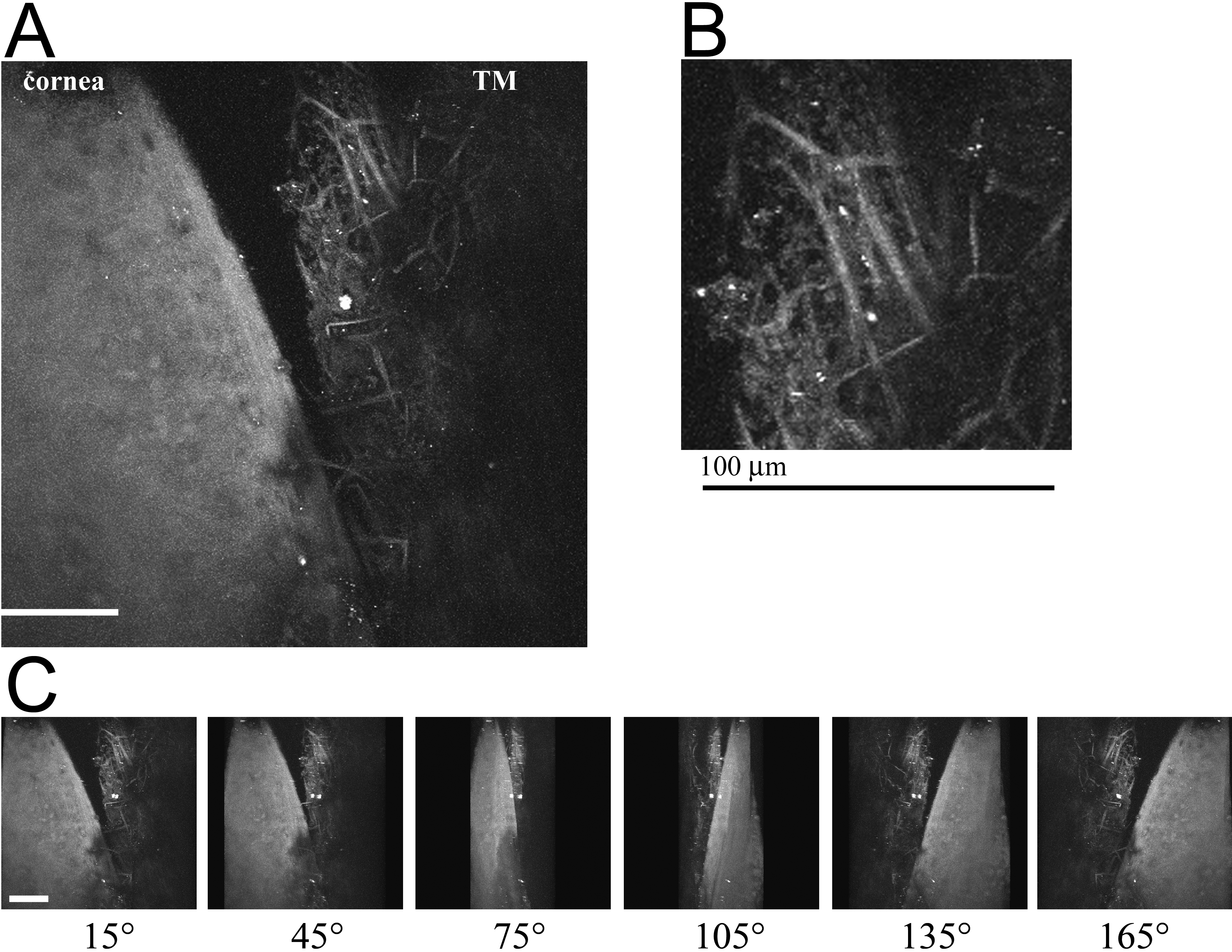
 Figure 1 of Ammar, Mol Vis 2010; 16:935-944.