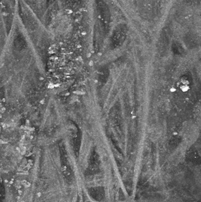
Figure 4 of Ammar, Mol Vis 2010; 16:935-944.


Figure 4. A 3D reconstruction of the TM region shown in Figure 3. The region of TM shown in Figure 3 was imaged by AF at 1 µm intervals. A total of 75 z-sections were projected into a 3D-animation with rotation about the y-axis. The AF signal from the collagen within the TM reveals a meshwork of collagen structures interwoven with what appears to be fluid spaces. The animation begins with a view of the aqueous face of the TM; rotation by 180 degrees reveals an open pore-like structure that penetrates the entire thickness scanned. The non-fibrous structure present at the aqueous surface can be viewed through the pore.

Note that the slide bar at the bottom of the quicktime movie can be used to manually control the flow of the movie, or double click on the image to automatically run the animation. If you are unable to view the movies, a representative frame from each movie is included below.


This animation requires Quicktime 6 or later.
Quicktime is available as a free download.