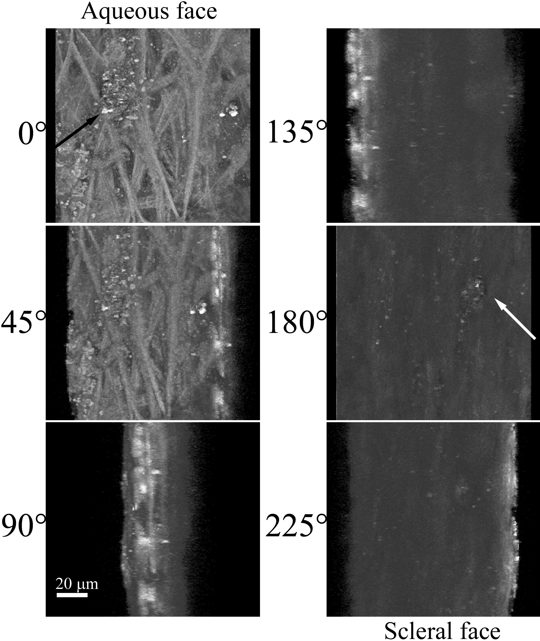
Figure 3. AF of the TM region of a human
eye from a 73-year-old donor. Another section TM tissue was
flat-mounted with the anterior chamber facing the microscope objective.
AF was collected as in
Figure 1. Single image snapshots
from the 3D reconstruction of the TM region are shown along with
corresponding degree of rotation (around the y-axis). 0° represents the
aqueous humor face of the TM, while 180° represents the
scleral-directed face. A non-fibrous structure present at the aqueous
surface (black arrow) can be viewed from the scleral face through an
open pore-like structure (white arrow). White scale bar=20 µm.
 Figure 3 of Ammar, Mol Vis 2010; 16:935-944.