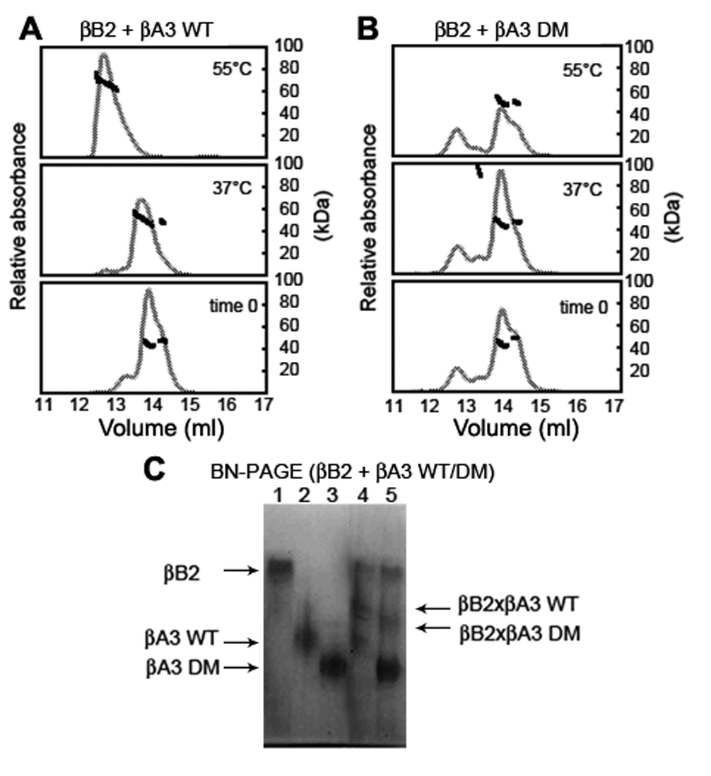
Figure 6. Hetero-oligomerization of βB2 with βA3. βB2 was mixed with either βA3 WT (
A) or βA3 DM (
B) and mixtures were immediately subjected to SEC-MALS (bottom panel) or incubated at 37 °C (middle panel) or 55 °C (top panel)
for 90 min as for βB1 in
Figure 3. Molar masses of βB2:βA3 WT peaks indicated predominantly dimers at 37 °C with a slight shift in elution volume and oligomers
at 55 °C. Molar masses of βB2:βA3 DM peaks indicated predominantly dimers at 37 °C without a shift in elution volume and no
oligomers were detected at 55 °C. Differences in hetero-oligomer formation were confirmed by subjecting mixtures of βB2 with
βA3 WT or βA3 DM incubated at 37 °C for 60 min to Blue-Native-PAGE (
C). Proteins were βB2 (lane 1), βA3 WT (lane 2), βA3 DM (lane 3), βB2:βA3 WT (lane 4), and βB2:βA3 DM (lane 5). Arrows indicate
migration position of proteins.
 Figure 6 of
Takata, Mol Vis 2009; 15:241-249.
Figure 6 of
Takata, Mol Vis 2009; 15:241-249.