Figure 6. Morphology of cells used for collection of data in
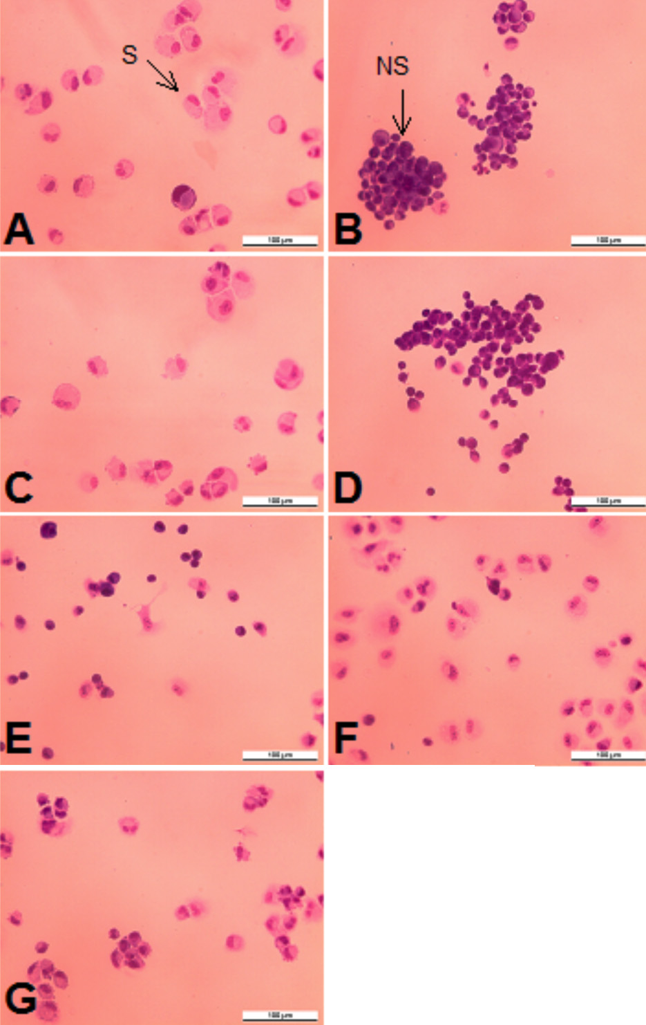
Figure 7. Representative light microscopic fields are shown. (
A) Untreated RPE cells grown on Fn; (
B) after preincubation with 250 μg/ml Gal-1 on Fn; (
C) Untreated RPE cells grown on Lam; (
D) after preincubation with 250 ug/ml Gal-1 on Lam; (
E) Untreated RPE cells grown on Gal-1; (
F) on Gal-1 and Fn; (
G) on Gal-1 and Lam. Spreading cells (S) were defined as cells with cytoplasmic protrusions and perinuclear halo formation,
and non spreading cells (NS) as rounded cells with little cytoplasmic spreading. The photographs represent a 100 fold magnification.
 Figure 6 of
Alge-Priglinger, Mol Vis 2009; 15:2162-2173.
Figure 6 of
Alge-Priglinger, Mol Vis 2009; 15:2162-2173.