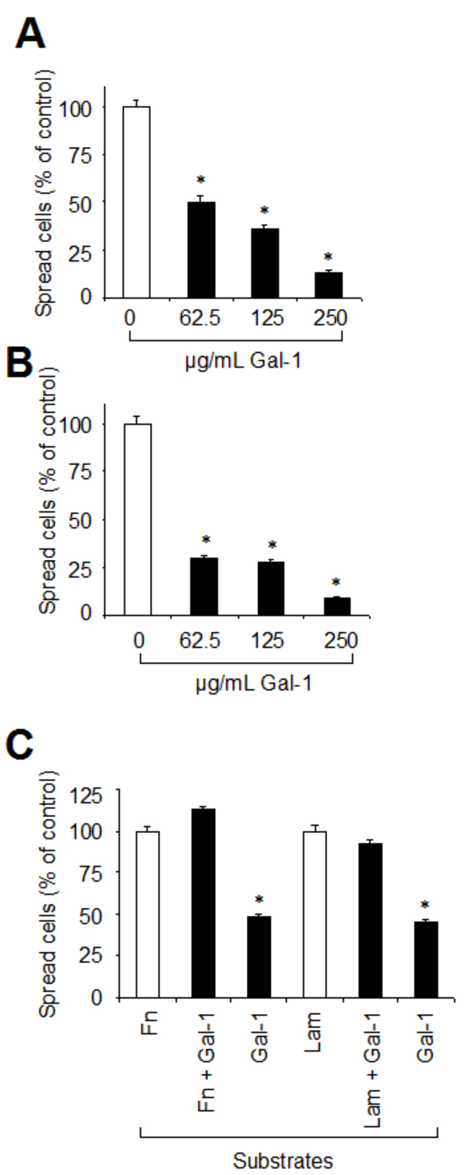
Figure 7. Galectin-1 inhibits spreading of RPE cells on fibronectin and laminin. A, B: RPE suspensions were incubated for 35 min with the indicated concentrations of galectin-1 and then plated on four chamber
slides coated with either Fn (A) or Lam (B) at a density of 5×10⁴ cells per chamber. Cells were allowed to spread in DMEM for 3.5 h at 37 °C before fixation and Giemsa staining. Values indicate
means±SD of four experiments performed in duplicate and are expressed as percentage of controls without galectin-1 in the
medium. (C) RPE cell spreading on galectin-1 as a substrate. Four chamber slides were coated with either Fn alone, Fn+galectin-1 (Gal-1),
Lam alone, Lam+Gal-1, or Gal-1 alone. RPE cells were then plated at a density of 5×10⁴ cells per chamber and allowed to spread. Results were obtained from evaluation of four separate fields by examining at least
100 cells per field. Values indicate means±SD of three experiments performed in duplicate and are expressed as percentage
of cells spreading on Fn or Lam as a substrate. Statistical analysis was performed using Students t-test and a p value <0.05 was considered as statistically significant (*).

 Figure 7 of
Alge-Priglinger, Mol Vis 2009; 15:2162-2173.
  Figure 7 of
Alge-Priglinger, Mol Vis 2009; 15:2162-2173.