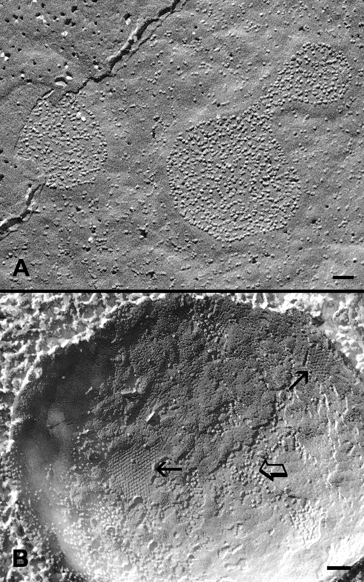
Figure 4. High pressure freezing of gap
junctions in cortical fiber cells. Freeze-fracture TEM in conjunction
with high pressure freezing shows that while gap junctions display only
loosely-packed connexons in superficial cortical fibers (
A), a
mixture of crystalline-packed (arrows) and loosely-arranged (opened
arrow) connexons can be visualized in the deeper cortical fibers of the
lens without prior chemical fixation (
B). This additional
experimental approach is to confirm that the structural remodeling of
gap junctions as observed in the chemically fixed lenses in
Figure 2
and
Figure 3
is a real change during fiber cell maturation. The scale bars indicate
100 nm.
 Figure 4 of Biswas, Mol Vis 2009; 15:1492-1508.