Figure 2. Gap junctions in the outer young
cortical fibers (0-400 μm from the surface). A: Both small
(arrows) and newly-formed gap junctions (opened arrows) in very
superficial cells display loosely-packed connexons. B & C:
Several larger gap junction plaques in slightly deeper cells also
exhibit loosely-packed connexons. In the images, pf is the P face of
the membrane and ef is the E face of the membrane. The scale bars
indicate 200 nm.
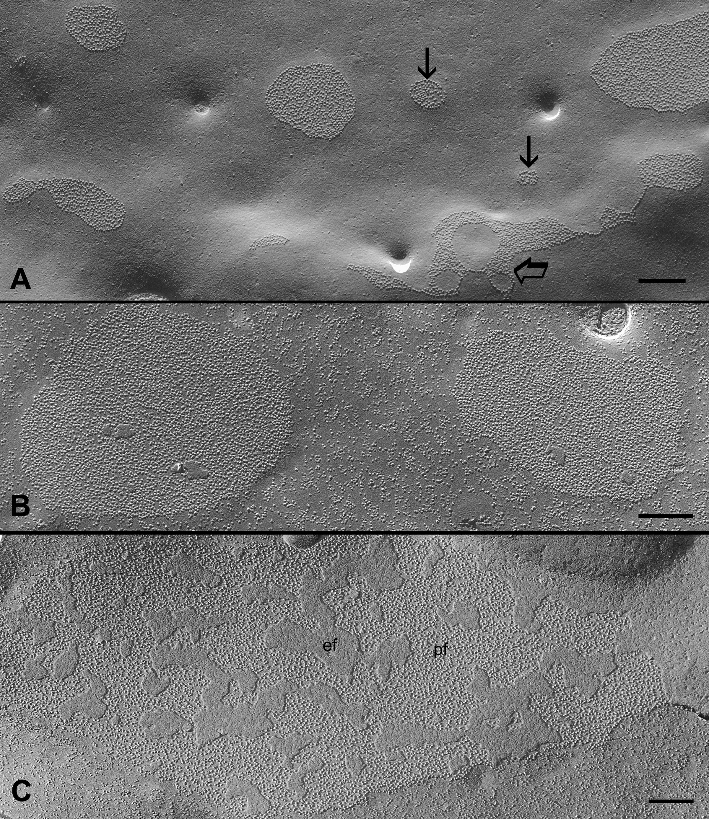
 Figure 2 of Biswas, Mol Vis 2009; 15:1492-1508.