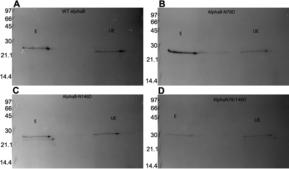
Figure 7. Two-dimensional protein profile of UV-A-exposed and unexposed WT αB and its three deamidated mutant proteins.
A-D show UV-A-exposed (E) and UV-A unexposed (UE) 2D-gel electrophoretic profiles of WT αB-crystallin and its three deamidated
mutant proteins. The unexposed and exposed preparations from each of the proteins were first separated by IEF in the first
dimension and then by SDS–PAGE in the second dimension. Four spots from each profile were identified (see
Figure 8) and were analyzed by the MALDI-TOF mass spectrometric method.
 Figure 7 of
Mafia, Mol Vis 2008; 14:234-248.
Figure 7 of
Mafia, Mol Vis 2008; 14:234-248.