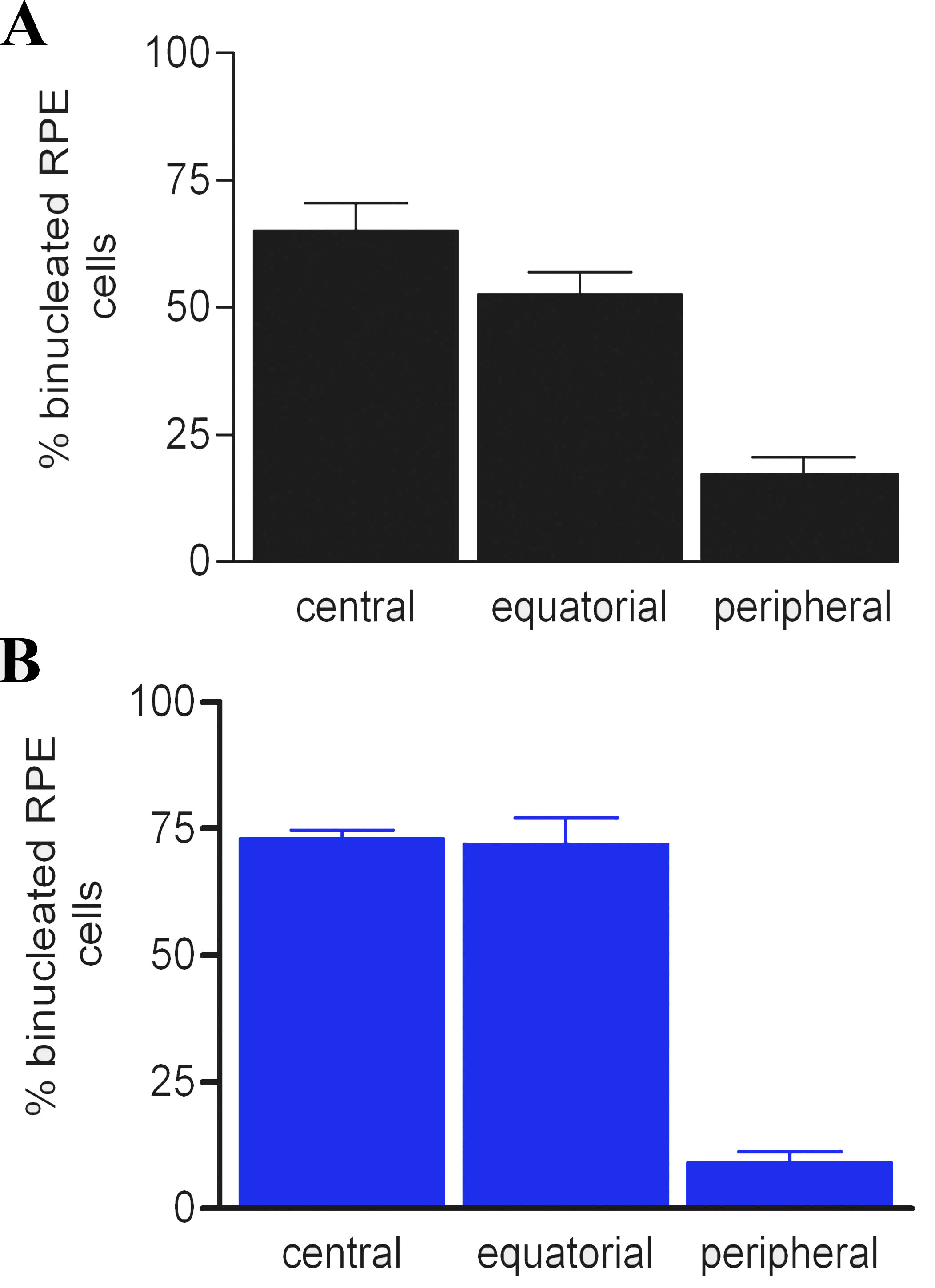
Figure 4. The percentage of binucleated
RPE cells in different retinal regions in pigmented (black bars) and
albino (blue bars) rats. In adult rodent retinas, many of the RPE cells
were binucleated. The proportion of these have been determined in both
pigmented (
A) and albino (
B) retinas. The retinas were
divided into three roughly equal geographic regions: central,
equatorial, and peripheral. In both pigmentation phenotypes the
majority of the binucleated cells were located toward the central
retina, although many were also found in equatorial regions. The
distribution of these cells is the reverse pattern of that found for
Ki67-positive cells shown in
Figure 2. The differences in
A
and
B are statistically significant (ANOVA, p<0.001). In
both cases, the differences between the percentage of binucleated RPE
cells between central and equatorial regions were not statistically
significant. However, differences in the percentage of binucleated RPE
cells found central and peripheral regions were significantly different
(Newman-Keuls p<0.01). The differences between equatorial and
peripheral regions were also statistically significant (Newman-Keuls
p<0.01).