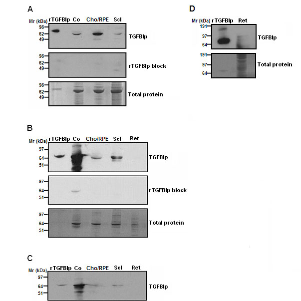
Figure 8. Transforming growth factor,
beta-induced protein detection in marmoset and human ocular tissues.
Western blot analysis identified the presence of transforming growth
factor, beta-induced protein (TGFBIp) in the cornea (Co), choroid/RPE
(Cho), sclera (Scl), and retina (Ret) of the marmoset (
A) and
human eye (
B). Pre-incubation of TGFBIp antibodies with
recombinant TGFBIp protein (rTGFBIp; 1 µM final concentration)
abolished all major anti-TGFBIp immunoreactive bands in marmoset tissue
extracts, with the exception of a faint band in the human cornea lane
migrating at ≤ 65 kDa (
B, center panel; rTGFBIp block). Total
protein loaded in each well was visualized by Coomassie Blue staining (
A
and
B, bottom panels).
C: Lighter exposure of
Figure 7B
(top panel). Recombinant TGFBIp protein (rTGFBIp, 40 ng) and cornea
served as positive controls.