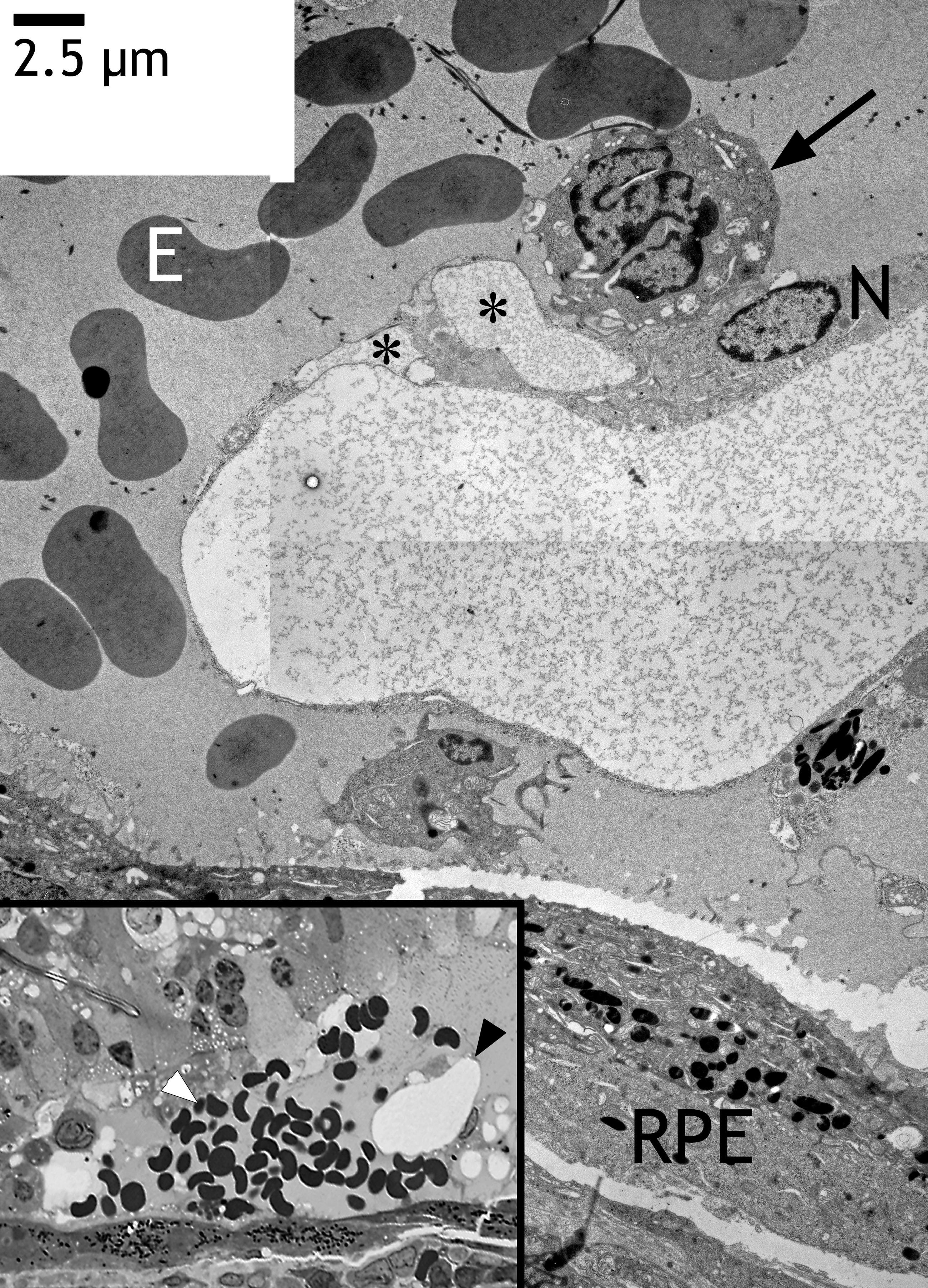
Figure 7. Newly formed capillary in the
subretinal space after transduction with HC Ad.VEGF-A from the same eye
as shown in
Figures
2J,K. The inset (left bottom) shows a semithin section with
a new capillary (black arrowhead) within the subretinal space and
erythrocytes originating probably from subretinal bleeding (white
arrowhead). The photoreceptors of the retina have degenerated and were
no longer visible. Electron microscopy revealed the same capillary
close to the level of the semithin section. The endothelium was very
thin, lacking extracellular matrix. However, it contained vacuoles
(asterisks). The endothelium did not contain any fenestrations, and the
lumen was free of erythrocytes (E). Note the extremely frayed or
fragmented nucleus of an endothelial cell (arrow). The nucleus (N) of
the endothelial cell that formed the vessel tube appeared normal. The
RPE formed two layers (bottom right).