![]() Figure 5 of
Johnson, Mol Vis 2007;
13:887-919.
Figure 5 of
Johnson, Mol Vis 2007;
13:887-919.
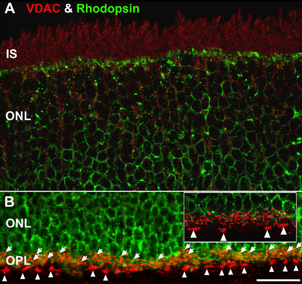
Figure 5. High magnification confocal images of outer retina double labeled for VDAC (red) and rhodopsin (green)
A: To reveal the voltage-dependent anion channel (VDAC) labeling of the IS (Figure 2B) the prominent rhodopsin labeling in the OSs and most IS was removed using Photoshop. Minimal colocalization of VDAC and rhodopsin occur in the ONL, highlighting the lack of mitochondria in most rod somas. However, the Müller glial cell processes that surround the rod somas are VDAC positive (data not shown). B: Stratification of rod spherule and cone pedicle mitochondria in the OPL. Double labeling for VDAC and rhodopsin shows the distal-proximal stratification of rod spherules (white arrows) and cone pedicles and their associated mitochondria. Tiers of rod spherules, each with one large mitochondrion, overly the rhodopsin-positive region. The more proximal cone pedicles contain clusters of mitochondria (white arrowheads) and are located in the rhodopsin-negative region. The inset is a higher magnification (2X) view of the OPL. Scale bar equal 20 μm for both panels.