![]() Figure 2 of
Johnson, Mol Vis 2007;
13:887-919.
Figure 2 of
Johnson, Mol Vis 2007;
13:887-919.
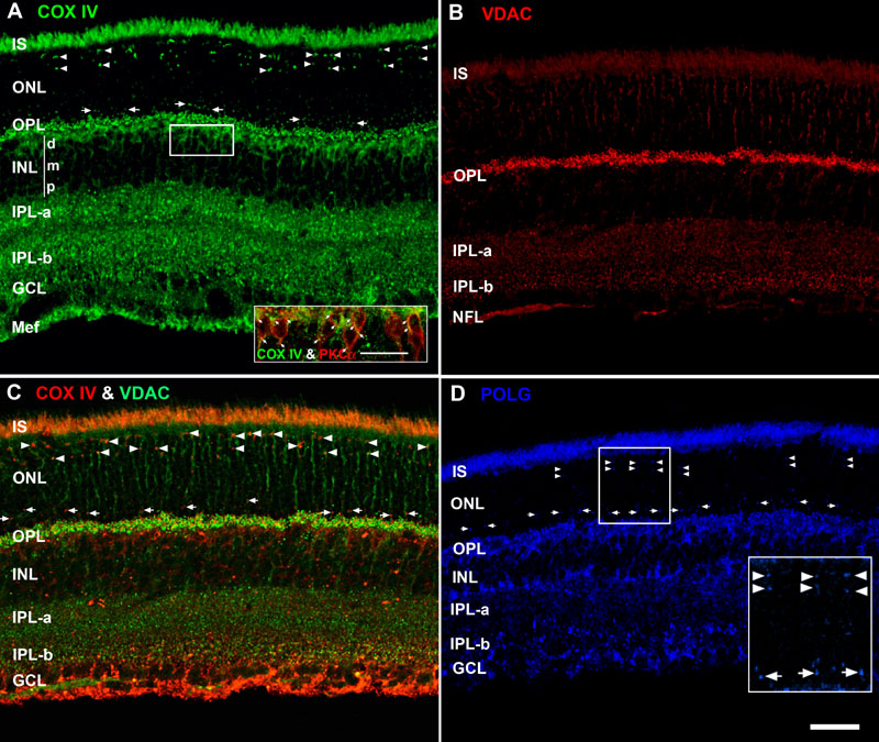
Figure 2. Molecular markers for three separate mitochondrial compartments reveal distinct retinal distribution and lamination patterns
The abbreviations of the retinal layers are used for this and all subsequent figures. IS represents inner segments, ONL represents outer nuclear layer, OPL represents outer plexiform layer, INL represents inner nuclear layer (d: distal, m: middle, p: proximal), IPL-α represents inner plexiform layer sublamina-α (OFF lamina), IPL-β: inner plexiform layer sublamina-β (ON lamina), GCL: ganglion cell layer, and Mef: Müller glial end-feet. A: Confocal image of retina immunolabeled for COX IV. The pairs of white arrowheads identify numerous intensely labeled arc-shaped puncta located in the distal ONL, which are juxtanuclear mitochondria in the cone somas. Between the white arrows, in the proximal ONL, is a band of small circular puncta that are juxtanuclear mitochondria near rod nuclei. The inset shows colocalization of COX IV (green) and PKCα (red) in rod bipolar cells (white arrows): scale bar equal 20 μm. B: Confocal image of retina immunostained for VDAC. Note the intense labeling in the OPL. C: Confocal image of retina double labeled with antibodies against COX IV (red) and VDAC (green). The pseudocoloring in panels A and B was reversed in this panel to clearly show the colocalization (yellow-orange pixels) in the IS, cone juxtanuclear mitochondria (white arrowheads) and rod juxtanuclear mitochondria (white arrows). Punctate colocalization also is present throughout the OPL, IPL-α, and IPL-β. D: Confocal image of retina immunolabeled for POLG. POLG also labels the cone juxtanuclear mitochondria (white arrowheads) and rod juxtanuclear mitochondria (white arrows). The inset is a higher magnification (2X) view. Scale bar equal 40 μm for all panels.