![]() Figure 11 of
Johnson, Mol Vis 2007;
13:887-919.
Figure 11 of
Johnson, Mol Vis 2007;
13:887-919.
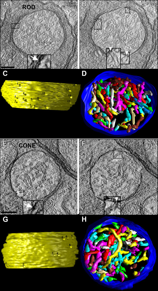
Figure 11. Three-dimensional electron tomograms of a rod spherule and a cone pedicle mitochondrion
A-D: Three-dimensional imaging of a rod spherule mitochondrion. A: A representative 2.2 nm slice through a tomographic volume of a rod spherule (from Figure 9A) that shows a large mitochondrion with many cristae. The inset (enlarged 3X) illustrates an example of a classical contact site, defined as the location where the OMM and IBM join (arrow). Scale bar for A and B equal 200 nm. B: Another slice through the volume showing two crista junctions (CJs: boxed) that have tubular openings, which connect the cristae with the intermembrane space. The inset at the bottom shows the openings (white arrowheads) of the two CJs enlarged 2X. C: Side view of the inner membrane of the segmented volume displayed with left lighting. CJ openings are invariably narrow, tubular and remarkably uniform in diameter. There are fifteen numbered CJ openings in this view. D: Top view of the segmented volume showing the outer membrane (blue) and a subset of cristae (various colors). Most of the cristae are tubular. However, some cristae possess lamellar compartments, which connect to the intermembrane space via CJs. E-H: Three-dimensional imaging of cone pedicle mitochondrion. E: A representative 2.2 nm slice through a tomographic volume of a cone pedicle (from Figure 9B) that shows a typical medium- to large-sized mitochondrion typical at this terminal. As illustrated, the abundance of cristae membranes is significantly smaller in each cone pedicle, compared to rod spherule, mitochondria. The inset (enlarged 3X) illustrates an example of a classical contact site (arrow). Scale bar for E and F equal 200 nm. F: Another slice through the volume showing two CJs (boxed). The inset at the bottom shows the openings (white arrowhead) of the two CJs expanded 2X. The openings of these crista junctions are significantly smaller than their counterparts in the rod spherule mitochondria (Table 3). G: Side view of the inner membrane of the segmented volume displayed with left lighting. The eight CJ openings seen in this view are numbered. H: Top view of the segmented volume showing the outer membrane (blue) and a subset of cristae (various colors). Most of the cristae are tubular, as in the rod spherule mitochondrion, although a small proportion have lamellar compartments. The cristae surface area and volume are significantly smaller in cone pedicle, compared to rod spherule, mitochondria (B, C).