![]() Figure 9 of
Johnson, Mol Vis 2007;
13:887-919.
Figure 9 of
Johnson, Mol Vis 2007;
13:887-919.
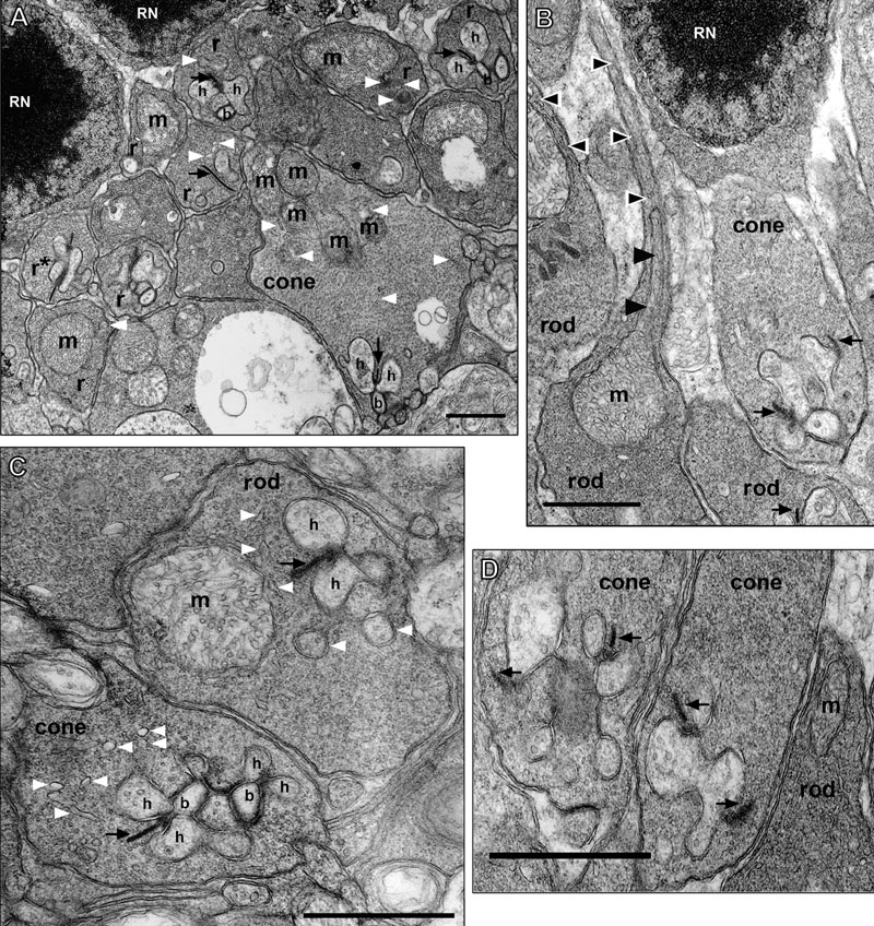
Figure 9. Ultrastructure of rod and cone photoreceptor synaptic terminals
A: The rod spherules (r) form two to four tiers that overly the larger more electron lucent cone pedicles (cone). Two rod nuclei (RN) are present. The rod spherules contain a single large mitochondrion (m), whereas the cone pedicles contain several mitochondria. Cisterna of smooth endoplasmic reticulum (white arrowheads) are often located adjacent to individual mitochondrion. Both terminals are filled with round synaptic vesicles. The invaginating processes of two lateral horizontal cells (h) and a central bipolar cell (b), the classic triad [122], are seen at the short synaptic ribbon (black arrow) of the cone and the longer synaptic ribbon (black arrow) of the rod in the upper right corner of the figure. The rod spherule on the left labeled r* appears to be a rod with two synaptic ribbon units as described [121]. B: Rod spherule and axon; cone pedicle. On the left portion of the panel, a descending rod axon (black and white arrowheads) and its expansion into a rod spherule (rod) is visible for two rods. An elongated mitochondrion (m; black arrowheads) is visible in the thin axon (0.20-0.25 μm) as it enlarges into the spherule. The rod spherules are filled with 25-35 nm electron dense round synaptic vesicles and a single large mitochondrion. A tangential view of an adjacent rod spherule with a synaptic ribbon (black arrow) is shown. A larger more electron lucent cone pedicle has two visible synaptic ribbons (black arrows) at invaginating synapses in this section. A rod nucleus (RN) in the most proximal row of the ONL is present. C: Rod spherule and cone pedicle. The rod spherules contain a single large mitochondrion (m) and an invaginating process with two lateral horizontal cells (h) and a central bipolar cell that is just out of the section. The rod has a long synaptic ribbon (black arrow) and a visible arciform density at its base. The most proximally located cone pedicle has two classic triads visible with two lateral horizontal cells (h) and a central bipolar cell (b). Other horizontal cell processes also are visible and the arciform density is evident. Cisterna of smooth endoplasmic reticulum (white arrowheads) are more numerous in cone pedicles than rod spherules. D: Two cone pedicles and a rod spherule. Each large cone pedicle has multiple synaptic ribbons (black arrows) at invaginating synapses. Several lateral horizontal cell processes, of varying size and intensity, are visible. The enlarged axonal region of rod spherule, with its darker matrix, contains a mitochondrion (m) that is just out of the section. Scale bars for all panels equal 1 μm.