![]() Figure 1 of
Johnson, Mol Vis 2007;
13:887-919.
Figure 1 of
Johnson, Mol Vis 2007;
13:887-919.
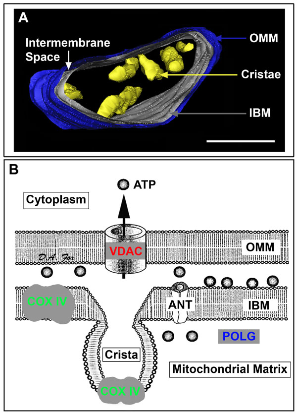
Figure 1. Rod inner segment mitochondrion and topographical markers of mitochondrial compartments
A: View of a mouse rod inner segment mitochondrion from a three-dimensional electron tomographic study after volume segmentation. The outer mitochondrial membrane (OMM) is blue, inner boundary membrane (IBM) apposed to the OMM is grey, cristae are yellow and a white arrow identifies the intermembrane space between the OMM and IBM. The IBM and cristal membranes form the contiguous, but distinct inner membrane system of the mitochondria [84,89,124,128]. Most cristae were removed graphically for illustrative purposes (adapted from [4]). Scale bar equals 200 nm. B: Schematic drawing of mitochondrial topology with site-specific compartments targeted for immunocytochemical experiments. Common immunocytochemical markers of the OMM (voltage-dependent anion channel: VDAC), inner membrane system (cytochrome oxidase IV: COX IV), and mitochondrial matrix (mitochondrial DNA polymerase-γ: POLG) are depicted. These mitochondrial compartments are consistent with the rod mitochondrion illustrated in A and the single label colorized confocal images shown in Figure 2A,B,D. Note the proximity of COX IV and VDAC to the adenine nucleotide transporter (ANT). The abbreviations COX IV, VDAC, and POLG are used in all subsequent figures.