![]() Figure 11 of
Hartung, Mol Vis 2007;
13:66-78.
Figure 11 of
Hartung, Mol Vis 2007;
13:66-78.
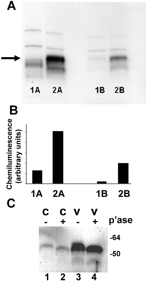
Figure 11. Vitreous increases c-fos protein expression and phosphorylation
A: Retinal pigment epithelial (RPE) cells were treated with 25% vitreous for one hour. Nuclear proteins were extracted and subjected to SDS-polyacrylamide gel electrophoresis and immunoblot analysis. Lanes 1A, 1B: Control nuclear protein extract. Lanes 2A, 2B: Nuclear protein extract from vitreous-treated RPE cells. Data shown are from two different RPE-vitreous donor pairs (A and B). The position of the c-fos band (62 kDa) is indicated by the arrow. B: Intensities of chemiluminescence corresponding to the bands at about 62 kDa in Figure 11A. A 4.3 fold (lane 2A compared to lane 1A) and 11.1 fold (lane 2B compared lane 1B) increase in c-fos protein was seen in the nuclear protein extracts of vitreous-treated RPE cells. C: RPE cells were treated with 25% vitreous for one hour. Nuclear proteins were extracted and treated with or without lambda protein phosphatase. Phosphatase (p'ase) treatment (+) resulted in the band at the position of c-fos moving faster in both control (lane 2) and vitreous-treated (lane 4) RPE nuclear protein extracts compared to nonphosphatase treated (-) samples (lanes 1, 3).