![]() Figure 5 of
Yaung, Mol Vis 2007;
13:566-577.
Figure 5 of
Yaung, Mol Vis 2007;
13:566-577.
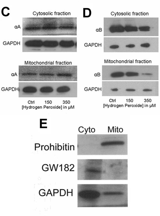
Figure 5. Mitochondrial distribution of α-crystallin protein in H2O2-treated retinal pigment epithelium
A and B represent quantitation of fluorescence in five experiments conducted in a similar fashion to that shown in Figure 3. Fractionated mitochondria and cytosolic proteins from H2O2-treated human retinal pigment epithelium (RPE) and controls were analyzed for α-crystallin content. Western blot analysis of αA-crystallin showed no apparent change in expression in either compartment with H2O2 exposure (C). αB-Crystallin in the cytosol remained relatively unchanged with varying doses of H2O2; however, mitochondrial α-crystallin decreased with increasing H2O2 dose (D). Purity of mitochondrial and cytosolic fractions was verified prior to crystallin analysis by two specific markers: prohibitin (E, top) and GM182 (E, middle). GAPDH, whose content is known to be much higher in cytoplasm than mitochondria, was also confirmed under our isolation conditions (E, bottom). Only traces (<1%) of inter-organelle contaminants were found. These fractions were then used for subsequent experiments. Asterisks indicate p<0.05 versus untreated controls. Cyto indicates cytosol; Mito indicates mitochondria.